uitsluitend voor onderzoeksdoeleinden
Tubastatin A HCl HDAC remmer
Cat.Nr.S2627
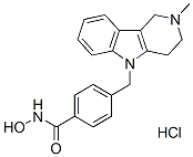
Chemische structuur
Moleculair gewicht: 371.86
Kwaliteitscontrole
Celkweek, behandeling & werkconcentratie
| Cellijnen | Assaytype | Concentratie | Incubatietijd | Formulering | Activiteitsbeschrijving | PMID |
|---|---|---|---|---|---|---|
| neuron cultures | Kinase assay | 2.5 μM | DMSO | induces α-tubulin hyperacetylation | ||
| neuron cultures | Function assay | ~10 μM | DMSO | protects against glutathione depletion-induced oxidative stress | ||
| 134/04 | Function assay | 7.5 µM | impairs myotube formation | |||
| C2C12 | Function assay | 7.5 µM | impairs myotube formation | |||
| HaCaT keratinocytes | Function assay | 10 μM | blocks arsenite from inducing Nrf2 protein translation | |||
| JURL-MK1 | Function assay | 10 μM | enhances cell adhesivity to fibronectin | |||
| CML-T1 | Function assay | 10 μM | enhances cell adhesivity to fibronectin | |||
| K562 | Function assay | 10 μM | enhances cell adhesivity to fibronectin | |||
| HL-60 | Function assay | 10 μM | enhances cell adhesivity to fibronectin | |||
| KMCH | Growth inhibitory assay | ~10 μM | decreases proliferation and anchorage-independent growth | |||
| THP-1 | Function assay | ~10 μM | inhibits TNF-α and IL-6 secretion | |||
| RAW 264.7 | Function assay | ~10 μM | attenuates NO production | |||
| HT3 | Function assay | ~5 μM | DMSO | induces the differential α-tubulin acetylation | ||
| SiHa | Function assay | ~5 μM | DMSO | induces the differential α-tubulin acetylation | ||
| CaSki | Function assay | ~5 μM | DMSO | induces the differential α-tubulin acetylation | ||
| SiHa | Function assay | ~5 μM | DMSO | inhibits Thapsigargin- or EGF-induced SOCE activation | ||
| CaSki | Function assay | ~5 μM | DMSO | inhibits Thapsigargin- or EGF-induced SOCE activation | ||
| MCF-7 | Growth inhibitory assay | 30 μM | DMSO | IC50=15 μM | ||
| MCF-7 | Function assay | 30 μM | DMSO | increases the microtubule acetylation level. | ||
| MCF-7 | Function assay | 30 μM | DMSO | stabilizes microtubules against cold-induced depolymerization | ||
| MCF-7 | Function assay | 15 μM | DMSO | stabilizes microtubules against nocodazole-induced disassembly | ||
| MCF-7 | Function assay | 30 μM | DMSO | alteres the assembly dynamics of interphase microtubules | ||
| MCF-7 | Function assay | 30 μM | DMSO | increases the binding of HDAC6 with interphase microtubules | ||
| PC12 | Function assay | ~3 μM | DMSO | up-regulates anti-oxidative gene expression related to transcription factor XBP1s | ||
| PC12 | Growth inhibitory assay | ~3 μM | DMSO | reverse H2O2-induced growth inhibition | ||
| HEK293T | Function assay | ~3 μM | DMSO | up-regulated XBP1s protein level | ||
| HEK293T | Function assay | ~3 μM | DMSO | delays XBP1s protein degradation via acetylation-mediated proteasomal degradation | ||
| Huh7 | Function assay | ~5 μM | DMSO | suppresses proliferation of hepatitis C virus replicon with EC50 = 0.3 μM | ||
| SKMEL21 | Growth inhibitory assay | ~500 nM | DMSO | inhibits cell proliferation | ||
| SKMEL103 | Growth inhibitory assay | ~500 nM | DMSO | inhibits cell proliferation | ||
| SKMEL28 | Growth inhibitory assay | ~500 nM | DMSO | inhibits cell proliferation | ||
| WM164 | Growth inhibitory assay | ~500 nM | DMSO | inhibits cell proliferation | ||
| WM1361a | Growth inhibitory assay | ~500 nM | DMSO | inhibits cell proliferation | ||
| WM1366 | Growth inhibitory assay | ~500 nM | DMSO | inhibits cell proliferation | ||
| WM793 | Growth inhibitory assay | ~500 nM | DMSO | inhibits cell proliferation | ||
| WM35 | Growth inhibitory assay | ~500 nM | DMSO | inhibits cell proliferation | ||
| WM983a | Growth inhibitory assay | ~500 nM | DMSO | inhibits cell proliferation | ||
| WM793 | Function assay | ~6 μM | DMSO | induce G1 arrest | ||
| WM164 | Function assay | ~6 μM | DMSO | induce G1 arrest | ||
| WM983a | Function assay | ~6 μM | DMSO | induce G1 arrest | ||
| WM164 | Function assay | ~3 μM | DMSO | augments expression of MHC class I and melanoma associated antigens | ||
| WM983a | Function assay | ~3 μM | DMSO | augments expression of MHC class I and melanoma associated antigens | ||
| IPC298 | Function assay | ~3 μM | DMSO | augments expression of MHC class I and melanoma associated antigens | ||
| SKMEL30 | Function assay | ~3 μM | DMSO | augments expression of MHC class I and melanoma associated antigens | ||
| TCa83 | Function assay | induces PTEN expression and membrane translocation | ||||
| 293T | Function assay | ~2 μg/ml | induces PTEN expression and membrane translocation | |||
| SACC-83 | Function assay | ~2 μg/ml | induces PTEN expression and membrane translocation | |||
| 293T | Function assay | ~2 μg/ml | induces PTEN acetylation at K163 | |||
| U-87 MG | Function assay | ~2 μg/ml | inhibits the migration and invasion | |||
| U-87 MG | Function assay | ~10 μM | inhibits AKT phosphorylation | |||
| U-87 MG | Growth inhibitory assay | ~10 μM | inhibits cell growth | |||
| Klik om meer experimentele gegevens over de cellijn te bekijken | ||||||
Chemische informatie, Opslag en Stabiliteit
| Moleculair gewicht | 371.86 | Formule | C20H21N3O2.HCl |
Opslag (Vanaf de ontvangstdatum) | |
|---|---|---|---|---|---|
| CAS-nr. | 1310693-92-5 | SDF downloaden | Opslag van stamoplossingen |
|
|
Oplosbaarheid
|
In vitro |
DMSO
: 100 mg/mL
(268.91 mM)
Verwarmd met 50°C waterbad;
Geultrasoneerd;
Water : Insoluble Ethanol : Insoluble |
Molariteitscalculator
|
In vivo |
|||||
In vivo Formuleringscalculator (Heldere oplossing)
Stap 1: Voer de onderstaande informatie in (Aanbevolen: Een extra dier voor het geval van verlies tijdens het experiment)
Stap 2: Voer de in vivo formulering in (Dit is alleen de calculator, geen formulering. Neem eerst contact met ons op als er geen in vivo formulering is in het gedeelte Oplosbaarheid.)
Berekeningsresultaten:
Werkconcentratie: mg/ml;
Methode voor het bereiden van DMSO-mastervloeistof: mg geneesmiddel vooraf opgelost in μL DMSO ( Concentratie mastervloeistof mg/mL, Neem eerst contact met ons op als de concentratie de DMSO-oplosbaarheid van de partij geneesmiddel overschrijdt. )
Methode voor het bereiden van in vivo formulering: Neem μL DMSO mastervloeistof, voeg vervolgens toeμL PEG300, mengen en helder maken, voeg vervolgens toeμL Tween 80, mengen en helder maken, voeg vervolgens toe μL ddH2O, mengen en helder maken.
Methode voor het bereiden van in vivo formulering: Neem μL DMSO mastervloeistof, voeg vervolgens toe μL Maïsolie, mengen en helder maken.
Opmerking: 1. Zorg ervoor dat de vloeistof helder is voordat u het volgende oplosmiddel toevoegt.
2. Zorg ervoor dat u het/de oplosmiddel(en) in de juiste volgorde toevoegt. U moet ervoor zorgen dat de verkregen oplossing, bij de vorige toevoeging, een heldere oplossing is voordat u verdergaat met het toevoegen van het volgende oplosmiddel. Fysische methoden zoals vortexen, echografie of een warmwaterbad kunnen worden gebruikt om het oplossen te bevorderen.
Werkingsmechanisme
| Targets/IC50/Ki |
HDAC6
(Cell-free assay) 15 nM
HDAC8
(Cell-free assay) 854 nM
|
|---|---|
| In vitro |
Tubastatin A is aanzienlijk selectief voor alle 11 HDAC-isoformen en behoudt een meer dan 1000-voudige selectiviteit tegen alle isovormen met uitzondering van HDAC8, waar het ongeveer 57-voudige selectiviteit heeft. In homocysteïnezuur (HCA) geïnduceerde neurodegeneratietests vertoont Tubastatin A een dosisafhankelijke bescherming tegen HCA-geïnduceerde neuronale celdood, beginnend bij 5 μM met bijna volledige bescherming bij 10 μM. Bij 100 ng/mL verhoogt Tubastatin A de onderdrukking van T-celproliferatie in vitro door Foxp3+ T-regulerende cellen (Tregs). Tubastatin A-behandeling in C2C12-cellen zou leiden tot een verminderde myotube-vorming wanneer alfa-tubuline vroeg in het myogene proces hypergeacetyleerd is; echter, myotube-elongatie treedt op wanneer alfa-tubuline hypergeacetyleerd is in myotubes. Een recente studie geeft aan dat Tubastatin A-behandeling de celelasticiteit verhoogt, zoals gebleken uit atoomkrachtmicroscopie (AFM) tests, zonder drastische veranderingen teweeg te brengen in de actine-microfilament- of microtubule-netwerken in muizenovariumkankercellijnen, MOSE-E en MOSE-L.
|
| Kinase Assay |
Enzyme-inhibitieassays
|
|
Enzymremmingsassays worden uitgevoerd door de Reaction Biology Corporation, Malvern, PA, met behulp van het Reaction Biology HDAC Spectrum-platform. (www.reactionbiology.com) De HDAC1, 2, 4, 5, 6, 7, 8, 9, 10 en 11 assays gebruiken geïsoleerd recombinant menselijk eiwit; HDAC3/NcoR2-complex wordt gebruikt voor de HDAC3-assay. Substraat voor HDAC1, 2, 3, 6, 10 en 11 assays is een fluorogene peptide van p53-residuen 379-382 (RHKKAc); substraat voor HDAC8 is een fluorogene diacylpeptide gebaseerd op residuen 379-382 van p53 (RHKAcKAc). Acetyl-Lys (trifluoracetyl)-AMC-substraat wordt gebruikt voor HDAC4, 5, 7 en 9 assays. Tubastatin A wordt opgelost in DMSO en getest in 10-dosis IC50-modus met 3-voudige seriële verdunning beginnend bij 30 μM. Controleverbinding Trichostatin A (TSA) wordt getest in een 10-dosis IC50 met 3-voudige seriële verdunning beginnend bij 5 μM. IC50-waarden worden geëxtraheerd door de dosis/respons-hellingen te curvenpassen.
|
|
| In vivo |
Dagelijkse behandeling met Tubastatin A van 0,5 mg/kg remt HDAC6 om de onderdrukkende activiteit van Tregs te bevorderen in muismodellen van ontsteking en auto-immuniteit, inclusief meerdere vormen van experimentele colitis en volledig major histocompatibility complex (MHC)-incompatibele cardiale allograft-afstoting.
|
Referenties |
|
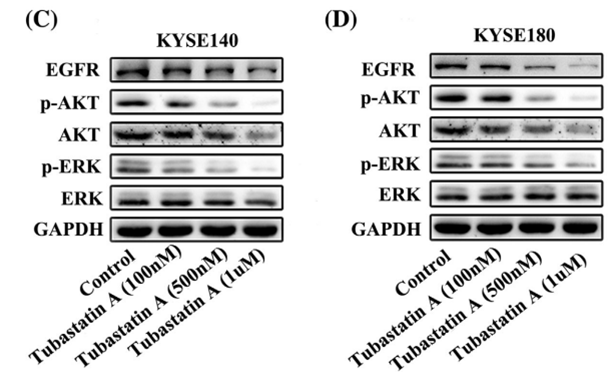
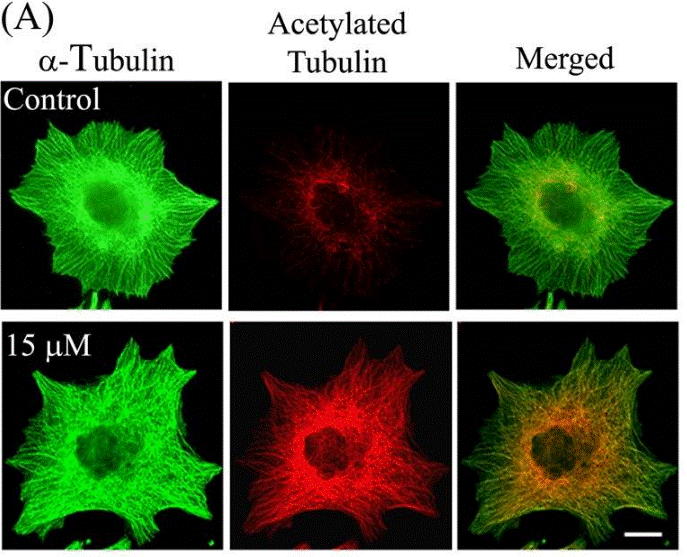
Toepassingen
| Methoden | Biomarkers | Afbeeldingen | PMID |
|---|---|---|---|
| Western blot | EGFR / p-AKT / AKT / p-ERK / ERK |
|
29665050 |
| Immunofluorescence | α-tubulin / Acetylated tubulin HDAC6 |
|
23798680 |
Technische ondersteuning
Tel: +1-832-582-8158 Ext:3
Als u nog andere vragen heeft, kunt u een bericht achterlaten.
Producten zijn uitsluitend voor onderzoeksdoeleinden. Niet voor menselijk gebruik. Wij verkopen niet aan patiënten.
©Copyright 2013 Selleck Chemicals. Alle rechten voorbehouden.