|
Comment citer 1. Pour la citation dans le texte (Matériel & Méthodes) : 2. Pour le tableau des ressources clés : |
||
|
Numéro vert : (877) 796-6397 -- États-Unis et Canada uniquement -- |
Fax : +1-832-582-8590 Commandes : +1-832-582-8158 |
Support technique : +1-832-582-8158 Ext:3 Veuillez indiquer votre numéro de commande dans le-mail. Nous nous efforçons de répondre à toutes les demandes par e-mail dans un délai dun jour ouvrable. |
Description biologique
| Spécificité | TRAF6 Antibody [B8B10] détecte les niveaux endogènes de la protéine TRAF6 totale. Cet anticorps ne devrait pas réagir de manière croisée avec d'autres membres de la famille TRAF. |
|---|---|
| Contexte | Les facteurs associés au récepteur du facteur de nécrose tumorale (TRAFs) constituent une famille de protéines adaptatrices cytoplasmiques qui médiatisent divers événements de signalisation intracellulaire. Chez les mammifères, sept membres de la famille TRAF ont été identifiés, dont six membres classiques (TRAF1–TRAF6) et un membre non classique (TRAF7). Les TRAFs classiques partagent un domaine TRAF carboxy-terminal conservé, qui est absent chez le TRAF7 non classique. Fonctionnellement, les TRAFs participent à la transduction du signal de la superfamille du facteur de nécrose tumorale (TNFSF) ainsi qu'aux membres de la superfamille des récepteurs Toll-like/IL-1 (TLR/ILR), régulant ainsi les cascades de signalisation en aval telles que la voie de la protéine kinase activée par les mitogènes (MAPK). Au-delà de la signalisation des récepteurs, les TRAFs jouent un rôle essentiel dans la prolifération cellulaire, la différenciation, l'apoptose et la survie, tout en modulant également les réponses immunitaires et inflammatoires. La dérégulation des protéines TRAF contribue à l'oncogenèse, avec des membres distincts exerçant soit des fonctions oncogènes, soit des fonctions de suppression tumorale. TRAF1, TRAF2, TRAF4, TRAF5 et TRAF6 ont été impliqués dans la carcinogenèse, tandis que TRAF3 fonctionne principalement comme un suppresseur tumoral. Parmi la famille, TRAF6 se distingue par sa spécificité de liaison aux récepteurs unique, lui permettant d'interagir non seulement avec les membres de la superfamille des récepteurs TNF, mais également avec les protéines de la superfamille IL-1R/TLR. Une surexpression de TRAF6 a été rapportée dans plusieurs types de tumeurs, y compris les carcinomes du côlon, gastriques et mammaires, ainsi que le mélanome. Fonctionnellement, TRAF6 favorise l'initiation et la progression tumorale en modulant l'apoptose, la prolifération, la survie et l'invasion. Mécaniquement, TRAF6 active plusieurs voies de signalisation, la cascade du récepteur Toll-like 4 (TLR4) étant particulièrement significative. Par des voies dépendantes et indépendantes de MyD88, TRAF6 amplifie la signalisation inflammatoire et de survie. De plus, TRAF6 améliore l'activation de la voie PI3K–AKT en facilitant l'ubiquitination de PI3K, augmentant ainsi la phosphorylation d'AKT et stimulant la croissance cellulaire. Collectivement, ces découvertes mettent en évidence TRAF6 comme un adaptateur critique qui intègre la signalisation des récepteurs avec les processus oncogènes. |
Informations dutilisation
| Application | WB, IP | Dilution |
|
||||
|---|---|---|---|---|---|---|---|
| Réactivité | Human, Monkey | ||||||
| Source | Rabbit Monoclonal Antibody | MW | 60 kDa | ||||
| Tampon de stockage | PBS, pH 7.2+50% Glycerol+0.05% BSA+0.01% NaN3 | Stockage (À partir de la date de réception) |
-20°C (avoid freeze-thaw cycles), 2 years | ||||
| WB |
Experimental Protocol:
Sample preparation
1. Tissue: Lyse the tissue sample by adding an appropriate volume of ice-cold RIPA/NP-40 Lysis Buffer (containing Protease Inhibitor Cocktail),and homogenize the tissue at a low temperature. 2. Adherent cell: Aspirate the culture medium and wash the cells with ice-cold PBS twice. Lyse the cells by adding an appropriate volume of RIPA/NP-40 Lysis Buffer (containing Protease Inhibitor Cocktail) and put the sample on ice for 5 min. 3. Suspension cell: Transfer the culture medium to a pre-cooled centrifuge tube. Centrifuge and aspirate the supernatant. Wash the cells with ice-cold PBS twice. Lyse the cells by adding an appropriate volume of RIPA/NP-40 Lysis Buffer (containing Protease Inhibitor Cocktail) and put the sample on ice for 5 min. 4. Place the lysate into a pre-cooled microcentrifuge tube. Centrifuge at 4°C for 15 min. Collect the supernatant;
5. Remove a small volume of lysate to determine the protein concentration;
6. Combine the lysate with protein loading buffer. Boil 20 µL sample under 95-100°C for 5 min. Centrifuge for 5 min after cool down on ice.
Electrophoretic separation
1. According to the concentration of extracted protein, load appropriate amount of protein sample and marker onto SDS-PAGE gels for electrophoresis. Recommended separating gel (lower gel) concentration: 10%. Reference Table for Selecting SDS-PAGE Separation Gel Concentrations 2. Power up 80V for 30 minutes. Then the power supply is adjusted (110 V~150 V), the Marker is observed, and the electrophoresis can be stopped when the indicator band of the predyed protein Marker where the protein is located is properly separated. (Note that the current should not be too large when electrophoresis, too large current (more than 150 mA) will cause the temperature to rise, affecting the result of running glue. If high currents cannot be avoided, an ice bath can be used to cool the bath.)
Transfer membrane
1. Take out the converter, soak the clip and consumables in the pre-cooled converter;
2. Activate PVDF membrane with methanol for 1 min and rinse with transfer buffer;
3. Install it in the order of "black edge of clip - sponge - filter paper - filter paper - glue -PVDF membrane - filter paper - filter paper - sponge - white edge of clip"; 4. The protein was electrotransferred to PVDF membrane. ( 0.45 µm PVDF membrane is recommended ) Reference Table for Selecting PVDF Membrane Pore Size Specifications Recommended conditions for wet transfer: 200 mA, 120 min. ( Note that the transfer conditions can be adjusted according to the protein size. For high-molecular-weight proteins, a higher current and longer transfer time are recommended. However, ensure that the transfer tank remains at a low temperature to prevent gel melting.)
Block
1. After electrotransfer, wash the film with TBST at room temperature for 5 minutes;
2. Incubate the film in the blocking solution for 1 hour at room temperature;
3. Wash the film with TBST for 3 times, 5 minutes each time.
Antibody incubation
1. Use 5% skim milk powder to prepare the primary antibody working liquid (recommended dilution ratio for primary antibody 1:1000), gently shake and incubate with the film at 4°C overnight; 2. Wash the film with TBST 3 times, 5 minutes each time;
3. Add the secondary antibody to the blocking solution and incubate with the film gently at room temperature for 1 hour;
4. After incubation, wash the film with TBST 3 times for 5 minutes each time.
Antibody staining
1. Add the prepared ECL luminescent substrate (or select other color developing substrate according to the second antibody) and mix evenly;
2. Incubate with the film for 1 minute, remove excess substrate (keep the film moist), wrap with plastic film, and expose in the imaging system.
|
Références
|
Données dapplication
WB
Validé par Selleck
-
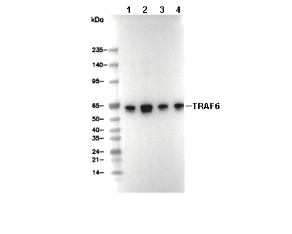 Lane 1: K562, Lane 2: Hela, Lane 3: 293T, Lane 4: COS-7