|
Comment citer 1. Pour la citation dans le texte (Matériel & Méthodes) : 2. Pour le tableau des ressources clés : |
||
|
Numéro vert : (877) 796-6397 -- États-Unis et Canada uniquement -- |
Fax : +1-832-582-8590 Commandes : +1-832-582-8158 |
Support technique : +1-832-582-8158 Ext:3 Veuillez indiquer votre numéro de commande dans le-mail. Nous nous efforçons de répondre à toutes les demandes par e-mail dans un délai dun jour ouvrable. |
Description biologique
| Spécificité | TCF12/HEB Antibody [G2F21] détecte les niveaux endogènes de la protéine TCF12/HEB totale. |
|---|---|
| Contexte | TCF12/HEB (Facteur de Transcription 12/Protéine de Liaison E-box de HeLa) est un facteur de transcription de classe I à hélice-boucle-hélice basique (bHLH) de la protéine E, encodé par TCF12 sur le chromosome 15q21, existant sous deux isoformes majeures : HEBCan (681 acides aminés, du promoteur ubiquitaire) et HEBAlt (forme courte, d'un promoteur distal). Ces isoformes hétérodimérisent avec des partenaires bHLH spécifiques aux tissus tels que E47, MyoD et NeuroD via leur domaine bHLH conservé, qui se compose d'une région basique de liaison à l'ADN et d'un motif HLH pour la dimérisation, afin de reconnaître les séquences consensus E-box CANNTG, pilotant ainsi l'engagement de la lignée dans les cellules T/B, les muscles et les neurones. TCF12/HEB contient un domaine d'activation N-terminal riche en résidus acides pour le recrutement de coactivateurs, un domaine de transactivation central avec des segments de glutamine/proline qui améliorent la pause et la libération de l'ARN polymérase II, le domaine bHLH central (~60 résidus) où l'hélice basique contacte le sillon majeur de l'ADN tandis que les hélices amphipathiques HLH dimérisent par des interfaces hydrophobes, et des domaines inhibiteurs C-terminaux qui modulent la spécificité des partenaires. L'épissage alternatif à l'exon 1 génère HEBCan pour les stades de développement des lymphocytes T et HEBAlt, qui est enrichi dans les thymocytes DN2/DN3 pour promouvoir une génération efficace de précurseurs. TCF12/HEB orchestre le développement des lymphocytes T en co-liant Lmo2 et Lyl1 aux amplificateurs Eβ et Cd4 pour activer les gènes impliqués dans la recombinaison et l'expansion, réprime les cibles E2A par dimérisation compétitive pour limiter l'auto-renouvellement, et équilibre la reconstitution des cellules souches hématopoïétiques (CSH) par rapport à la différenciation, une déficience entraînant un biais myéloïde, un blocage des cellules B/T et des défauts de prolifération. La signalisation PKCθ/Carma1 phosphoryle HEBCan sur des résidus sérine, levant l'autoinhibition médiée par la protéine Id et permettant la formation de boucles de chromatine induites par le TCR avec Runx1 et Foxp1 pour l'expression de Il2ra et Dtx1, tandis que HEBAlt s'associe uniquement à Bcl11b au niveau de l'amplificateur Tcrα pour faciliter la sélection positive. Les mutations de TCF12 provoquent une craniosynostose coronale (syndrome de Saethre-Chotzen) en perturbant l'hétérodimérisation de Twist1 et la perméabilité des sutures ; l'haploinsuffisance est liée à la dyslexie par dérégulation des gènes neurodéveloppementaux, et les altérations somatiques contribuent à la leucémie (fusions AML1-ETO) ou à la progression des tumeurs solides via l'EMT et l'invasion. |
Informations dutilisation
| Application | WB, IP, IHC | Dilution |
|
||||||
|---|---|---|---|---|---|---|---|---|---|
| Réactivité | Human | ||||||||
| Source | Rabbit Monoclonal Antibody | MW | 85 kDa | ||||||
| Tampon de stockage | PBS, pH 7.2+50% Glycerol+0.05% BSA+0.01% NaN3 | Stockage (À partir de la date de réception) |
-20°C (avoid freeze-thaw cycles), 2 years | ||||||
| WB |
Experimental Protocol:
Sample preparation
1. Tissue: Lyse the tissue sample by adding an appropriate volume of ice-cold RIPA/Nuclear Lysis Buffer (containing Protease Inhibitor Cocktail),and homogenize the tissue at a low temperature. 2. Adherent cell: Aspirate the culture medium and wash the cells with ice-cold PBS twice. Lyse the cells by adding an appropriate volume of RIPA/Nuclear Lysis Buffer (containing Protease Inhibitor Cocktail) and put the sample on ice for 5 min. 3. Suspension cell: Transfer the culture medium to a pre-cooled centrifuge tube. Centrifuge and aspirate the supernatant. Wash the cells with ice-cold PBS twice. Lyse the cells by adding an appropriate volume of RIPA/Nuclear Lysis Buffer (containing Protease Inhibitor Cocktail) and put the sample on ice for 5 min. 4. Place the lysate into a pre-cooled microcentrifuge tube. Centrifuge at 4°C for 15 min. Collect the supernatant;
5. Remove a small volume of lysate to determine the protein concentration;
6. Combine the lysate with protein loading buffer. Boil 20 µL sample under 95-100°C for 5 min. Centrifuge for 5 min after cool down on ice.
Electrophoretic separation
1. According to the concentration of extracted protein, load appropriate amount of protein sample and marker onto SDS-PAGE gels for electrophoresis. Recommended separating gel (lower gel) concentration: 10%. Reference Table for Selecting SDS-PAGE Separation Gel Concentrations 2. Power up 80V for 30 minutes. Then the power supply is adjusted (110 V~150 V), the Marker is observed, and the electrophoresis can be stopped when the indicator band of the predyed protein Marker where the protein is located is properly separated. (Note that the current should not be too large when electrophoresis, too large current (more than 150 mA) will cause the temperature to rise, affecting the result of running glue. If high currents cannot be avoided, an ice bath can be used to cool the bath.)
Transfer membrane
1. Take out the converter, soak the clip and consumables in the pre-cooled converter;
2. Activate PVDF membrane with methanol for 1 min and rinse with transfer buffer;
3. Install it in the order of "black edge of clip - sponge - filter paper - filter paper - glue -PVDF membrane - filter paper - filter paper - sponge - white edge of clip"; 4. The protein was electrotransferred to PVDF membrane. ( 0.45 µm PVDF membrane is recommended ) Reference Table for Selecting PVDF Membrane Pore Size Specifications Recommended conditions for wet transfer: 200 mA, 120 min. ( Note that the transfer conditions can be adjusted according to the protein size. For high-molecular-weight proteins, a higher current and longer transfer time are recommended. However, ensure that the transfer tank remains at a low temperature to prevent gel melting.)
Block
1. After electrotransfer, wash the film with TBST at room temperature for 5 minutes;
2. Incubate the film in the blocking solution for 1 hour at room temperature;
3. Wash the film with TBST for 3 times, 5 minutes each time.
Antibody incubation
1. Use 5% skim milk powder to prepare the primary antibody working liquid (recommended dilution ratio for primary antibody 1:1000), gently shake and incubate with the film at 4°C overnight; 2. Wash the film with TBST 3 times, 5 minutes each time;
3. Add the secondary antibody to the blocking solution and incubate with the film gently at room temperature for 1 hour;
4. After incubation, wash the film with TBST 3 times for 5 minutes each time.
Antibody staining
1. Add the prepared ECL luminescent substrate (or select other color developing substrate according to the second antibody) and mix evenly;
2. Incubate with the film for 1 minute, remove excess substrate (keep the film moist), wrap with plastic film, and expose in the imaging system.
|
| IHC |
Experimental Protocol:
Deparaffinization/Rehydration
1. Deparaffinize/hydrate sections:
2. Incubate sections in three washes of xylene for 5 min each.
3. Incubate sections in two washes of 100% ethanol for 10 min each.
4. Incubate sections in two washes of 95% ethanol for 10 min each.
5. Wash sections two times in dH2O for 5 min each.
6.Antigen retrieval: For Citrate: Heat slides in a microwave submersed in 1X citrate unmasking solution until boiling is initiated; continue with 10 min at a sub-boiling temperature (95°-98°C). Cool slides on bench top for 30 min.
Staining
1. Wash sections in dH2O three times for 5 min each.
2. Incubate sections in 3% hydrogen peroxide for 10 min.
3. Wash sections in dH2O two times for 5 min each.
4. Wash sections in wash buffer for 5 min.
5. Block each section with 100–400 µl of blocking solution for 1 hr at room temperature.
6. Remove blocking solution and add 100–400 µl primary antibody diluent in to each section. Incubate overnight at 4°C.
7. Remove antibody solution and wash sections with wash buffer three times for 5 min each.
8. Cover section with 1–3 drops HRPas needed. Incubate in a humidified chamber for 30 min at room temperature.
9. Wash sections three times with wash buffer for 5 min each.
10. Add DAB Chromogen Concentrate to DAB Diluent and mix well before use.
11. Apply 100–400 µl DAB to each section and monitor closely. 1–10 min generally provides an acceptable staining intensity.
12. Immerse slides in dH2O.
13. If desired, counterstain sections with hematoxylin.
14. Wash sections in dH2O two times for 5 min each.
15. Dehydrate sections: Incubate sections in 95% ethanol two times for 10 sec each; Repeat in 100% ethanol, incubating sections two times for 10 sec each; Repeat in xylene, incubating sections two times for 10 sec each.
16. Mount sections with coverslips and mounting medium.
|
Références
|
Données dapplication
WB
Validé par Selleck
-
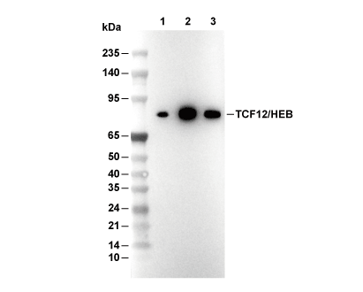 Lane 1: IMR-32, Lane 2: Jurkat, Lane 3: MOLT4