|
Comment citer 1. Pour la citation dans le texte (Matériel & Méthodes) : 2. Pour le tableau des ressources clés : |
||
|
Numéro vert : (877) 796-6397 -- États-Unis et Canada uniquement -- |
Fax : +1-832-582-8590 Commandes : +1-832-582-8158 |
Support technique : +1-832-582-8158 Ext:3 Veuillez indiquer votre numéro de commande dans le-mail. Nous nous efforçons de répondre à toutes les demandes par e-mail dans un délai dun jour ouvrable. |
Description biologique
| Spécificité | TCF11/NRF1 Antibody [F22N18] détecte les niveaux endogènes de la protéine TCF11/NRF1 totale. |
|---|---|
| Contexte | Le TCF11, également connu sous le nom de NRF1 (facteur nucléaire lié à l'érythroïde 2-), est un facteur de transcription CNC-bZIP (Cap'n'collar basic leucine zipper) appartenant à la famille Nrf, exprimé de manière ubiquitaire dans les tissus et existant sous plusieurs isoformes, y compris une forme de 120 kDa liée à la membrane du RE et une variante tronquée nucléaire de 65 kDa. Le TCF11 présente un domaine bZIP pour la liaison à l'ADN aux éléments de réponse antioxydante (AREs), Neh1L (domaine CNC avec bZIP), Neh2L (dégron ETGE/Neh2-like pour l'interaction avec Keap1), Neh4L/Neh5L (domaines de transactivation), et Neh6L (avec des motifs riches en sérine pour la phosphorylation de GSK3 et la dégradation médiée par CRL3), ainsi que des domaines N-terminaux acides sensibles au glucose et ciblant le RE qui permettent une répartition topologique. Le TCF11 se transloque du RE vers le noyau après inhibition du protéasome via une rétrotranslocation dépendante de p97/VCP et un traitement protéolytique, où il s'hétérodimérise avec de petites protéines Maf pour se lier aux AREs et induire transcriptionnellement les gènes des sous-unités du protéasome (par exemple, PSMB5-8), restaurant la capacité protéolytique et atténuant le stress protéotoxique. Ce mécanisme régule également les gènes de la biogenèse mitochondriale, les réponses au stress oxydatif via GCLC, et la cytoprotection contre les dommages induits par la roténone, le TCF11 exerçant des effets de répression tumorale plus forts que Nrf1α en régulant positivement les gènes de survie dans le carcinome hépatocellulaire. Une dérégulation relie le TCF11 à la neurodégénérescence et à la progression du cancer en raison d'une homéostase redox/protéostase altérée. |
Informations dutilisation
| Application | WB | Dilution |
|
||
|---|---|---|---|---|---|
| Réactivité | Human, Mouse, Monkey | ||||
| Source | Rabbit Monoclonal Antibody | MW | 120-140 kDa | ||
| Tampon de stockage | PBS, pH 7.2+50% Glycerol+0.05% BSA+0.01% NaN3 | Stockage (À partir de la date de réception) |
-20°C (avoid freeze-thaw cycles), 2 years | ||
| WB |
Experimental Protocol:
Sample preparation
1. Tissue: Lyse the tissue sample by adding an appropriate volume of ice-cold RIPA/NP-40 Lysis Buffer (containing Protease Inhibitor Cocktail),and homogenize the tissue at a low temperature. 2. Adherent cell: Aspirate the culture medium and wash the cells with ice-cold PBS twice. Lyse the cells by adding an appropriate volume of RIPA/NP-40 Lysis Buffer (containing Protease Inhibitor Cocktail) and put the sample on ice for 5 min. 3. Suspension cell: Transfer the culture medium to a pre-cooled centrifuge tube. Centrifuge and aspirate the supernatant. Wash the cells with ice-cold PBS twice. Lyse the cells by adding an appropriate volume of RIPA/NP-40 Lysis Buffer (containing Protease Inhibitor Cocktail) and put the sample on ice for 5 min. 5. Take a small amount of the lysate to determine the protein concentration; Electrophoretic separation
1. According to the concentration of extracted protein, load appropriate amount of protein sample and marker onto SDS-PAGE gels for electrophoresis. Recommended separating gel (lower gel) concentration: 5%. Reference Table for Selecting SDS-PAGE Separation Gel Concentrations 2. Power up 80V for 30 minutes. Then the power supply is adjusted (110 V~150 V), the Marker is observed, and the electrophoresis can be stopped when the indicator band of the predyed protein Marker where the protein is located is properly separated. (Note that the current should not be too large when electrophoresis, too large current (more than 150 mA) will cause the temperature to rise, affecting the result of running glue. If high currents cannot be avoided, an ice bath can be used to cool the bath.)
Transfer membrane
1. Take out the converter, soak the clip and consumables in the pre-cooled converter;
2. Activate PVDF membrane with methanol for 1 min and rinse with transfer buffer;
3. Install it in the order of "black edge of clip - sponge - filter paper - filter paper - glue -PVDF membrane - filter paper - filter paper - sponge - white edge of clip"; 4. The protein was electrotransferred to PVDF membrane. ( 0.45 µm PVDF membrane is recommended ) Reference Table for Selecting PVDF Membrane Pore Size Specifications Recommended conditions for wet transfer: 200 mA, 120 min. ( Note that the transfer conditions can be adjusted according to the protein size. For high-molecular-weight proteins, a higher current and longer transfer time are recommended. However, ensure that the transfer tank remains at a low temperature to prevent gel melting.)
Block
1. After electrotransfer, wash the film with TBST at room temperature for 5 minutes;
2. Incubate the film in the blocking solution for 1 hour at room temperature;
3. Wash the film with TBST for 3 times, 5 minutes each time.
Antibody incubation
1. Use 5% skim milk powder to prepare the primary antibody working liquid (recommended dilution ratio for primary antibody 1:1000), gently shake and incubate with the film at 4°C overnight; 2. Wash the film with TBST 3 times, 5 minutes each time;
3. Add the secondary antibody to the blocking solution and incubate with the film gently at room temperature for 1 hour;
4. After incubation, wash the film with TBST 3 times for 5 minutes each time.
Antibody staining
1. Add the prepared ECL luminescent substrate (or select other color developing substrate according to the second antibody) and mix evenly;
2. Incubate with the film for 1 minute, remove excess substrate (keep the film moist), wrap with plastic film, and expose in the imaging system.
|
Références
|
Données dapplication
WB
Validé par Selleck
-
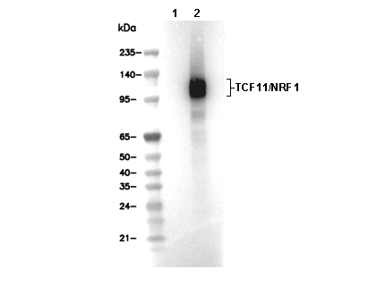 Lane 1: U-2 OS, Lane 2: U-2 OS (MG132, 10 µM, 8 h)