|
Comment citer 1. Pour la citation dans le texte (Matériel & Méthodes) : 2. Pour le tableau des ressources clés : |
||
|
Numéro vert : (877) 796-6397 -- États-Unis et Canada uniquement -- |
Fax : +1-832-582-8590 Commandes : +1-832-582-8158 |
Support technique : +1-832-582-8158 Ext:3 Veuillez indiquer votre numéro de commande dans le-mail. Nous nous efforçons de répondre à toutes les demandes par e-mail dans un délai dun jour ouvrable. |
Description biologique
| Spécificité | SET / TAF-I Antibody [N20C23] détecte les niveaux endogènes de la protéine SET/TAF-I totale. |
|---|---|
| Contexte | SET, également connu sous le nom de Template Activating Factor-I (TAF-I), est une protéine nucléaire exprimée de manière ubiquitaire dans les cellules somatiques de mammifères qui fonctionne comme une chaperonne d'histone H1 de liaison. Il existe sous deux isoformes, TAF-Iα et TAF-Iβ, qui sont encodées par le même locus génique mais transcrites à partir de promoteurs distincts, entraînant des modèles d'expression différentiels selon les types de cellules. Structurellement, TAF-I forme des homo- ou hétérodimères via un domaine de dimérisation central et possède une région C-terminale hautement acide essentielle à son activité de chaperonne d'histone. Bien que TAF-Iα et TAF-Iβ ne diffèrent que par leurs régions N-terminales, TAF-Iα présente une activité chaperonne plus faible en raison d'une interaction intramoléculaire auto-inhibitrice entre sa région N-terminale basique et sa queue C-terminale acide. Fonctionnellement, TAF-I régule la dynamique de l'histone H1 en facilitant à la fois l'assemblage et le désassemblage des chromatosomes, en modulant l'activité transcriptionnelle de manière spécifique au gène et au type de cellule, et en contribuant au remodelage de la chromatine et à l'intégrité du génome. |
Informations dutilisation
| Application | WB, IP, IHC, IF, FCM | Dilution |
|
||||||||||
|---|---|---|---|---|---|---|---|---|---|---|---|---|---|
| Réactivité | Human, Rat | ||||||||||||
| Source | Rabbit Monoclonal Antibody | MW | 33 kDa | ||||||||||
| Tampon de stockage | PBS, pH 7.2+50% Glycerol+0.05% BSA+0.01% NaN3 | Stockage (À partir de la date de réception) |
-20°C (avoid freeze-thaw cycles), 2 years | ||||||||||
| WB |
Experimental Protocol:
Sample preparation
1. Tissue: Lyse the tissue sample by adding an appropriate volume of ice-cold RIPA/NP-40 Lysis Buffer (containing Protease Inhibitor Cocktail),and homogenize the tissue at a low temperature or lyse it by sonication on ice, then incubate on ice for 30 minutes. 2. Adherent cell: Aspirate the culture medium and transfer the cells into an EP tube. Wash the cells with ice-cold PBS twice. Add an appropriate volume of RIPA/NP-40 Lysis Buffer (containing Protease Inhibitor Cocktail), sonicate to lyse the cells, and incubate on ice for 30 minutes. 3. Suspension cell: Transfer the culture medium to a pre-cooled centrifuge tube. Centrifuge and aspirate the supernatant. Wash the cells with ice-cold PBS twice.Add an appropriate volume of RIPA/NP-40 Lysis Buffer (containing Protease Inhibitor Cocktail), sonicate to lyse the cells, and incubate on ice for 30 minutes. 4. Place the lysate into a pre-cooled microcentrifuge tube. Centrifuge at 4°C for 15 min. Collect the supernatant;
5. Remove a small volume of lysate to determine the protein concentration;
6. Combine the lysate with protein loading buffer. Boil 20 µL sample under 95-100°C for 5 min. Centrifuge for 5 min after cool down on ice.
Electrophoretic separation
1. According to the concentration of extracted protein, load appropriate amount of protein sample and marker onto SDS-PAGE gels for electrophoresis. Recommended separating gel (lower gel) concentration: 10%. Reference Table for Selecting SDS-PAGE Separation Gel Concentrations 2. Power up 80V for 30 minutes. Then the power supply is adjusted (110 V~150 V), the Marker is observed, and the electrophoresis can be stopped when the indicator band of the predyed protein Marker where the protein is located is properly separated. (Note that the current should not be too large when electrophoresis, too large current (more than 150 mA) will cause the temperature to rise, affecting the result of running glue. If high currents cannot be avoided, an ice bath can be used to cool the bath.)
Transfer membrane
1. Take out the converter, soak the clip and consumables in the pre-cooled converter;
2. Activate PVDF membrane with methanol for 1 min and rinse with transfer buffer;
3. Install it in the order of "black edge of clip - sponge - filter paper - filter paper - glue -PVDF membrane - filter paper - filter paper - sponge - white edge of clip"; 4. The protein was electrotransferred to PVDF membrane. ( 0.45 µm PVDF membrane is recommended ) Reference Table for Selecting PVDF Membrane Pore Size Specifications Recommended conditions for wet transfer: 200 mA, 60 min. ( Note that the transfer conditions can be adjusted according to the protein size. For high-molecular-weight proteins, a higher current and longer transfer time are recommended. However, ensure that the transfer tank remains at a low temperature to prevent gel melting.)
Block
1. After electrotransfer, wash the film with TBST at room temperature for 5 minutes;
2. Incubate the film in the blocking solution for 1 hour at room temperature;
3. Wash the film with TBST for 3 times, 5 minutes each time.
Antibody incubation
1. Use 5% skim milk powder to prepare the primary antibody working liquid (recommended dilution ratio for primary antibody 1:1000000), gently shake and incubate with the film at 4°C overnight; 2. Wash the film with TBST 3 times, 5 minutes each time;
3. Add the secondary antibody to the blocking solution and incubate with the film gently at room temperature for 1 hour;
4. After incubation, wash the film with TBST 3 times for 5 minutes each time.
Antibody staining
1389. Add the prepared ECL luminescent substrate (or select other color developing substrate according to the second antibody) and mix evenly;
2. Incubate with the film for 1 minute, remove excess substrate (keep the film moist), wrap with plastic film, and expose in the imaging system.
|
Références
|
Données dapplication
IF
Validé par Selleck
-
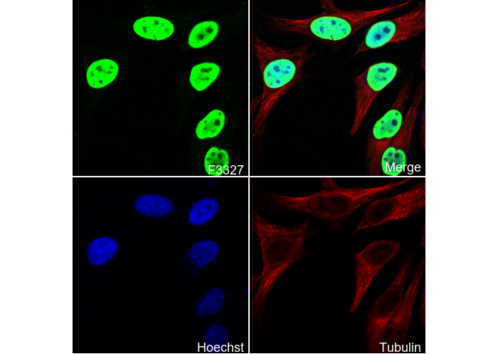 Immunofluorescent analysis of Hela cells using F3327 (green, 1:1000), Hoechst (blue) and tubulin (Red).
WB
Validé par Selleck
-
 Lane 1: Jurkat, Lane 2: 293T, Lane 3: Hela, Lane 4: HepG2