|
Comment citer 1. Pour la citation dans le texte (Matériel & Méthodes) : 2. Pour le tableau des ressources clés : |
||
|
Numéro vert : (877) 796-6397 -- États-Unis et Canada uniquement -- |
Fax : +1-832-582-8590 Commandes : +1-832-582-8158 |
Support technique : +1-832-582-8158 Ext:3 Veuillez indiquer votre numéro de commande dans le-mail. Nous nous efforçons de répondre à toutes les demandes par e-mail dans un délai dun jour ouvrable. |
Description biologique
| Spécificité | PSMD14 Antibody [G24K15] détecte les niveaux endogènes de la protéine PSMD14 totale. |
|---|---|
| Contexte | PSMD14, également connue sous le nom de sous-unité régulatrice non-ATPase 14 du protéasome 26S ou Rpn11, est une métalloenzyme critique au sein du couvercle de la particule régulatrice 19S (RP) du complexe protéasomique 26S et est un membre de la famille JAMM/MPN+ des déubiquitinases (DUBs). Elle présente un domaine MPN contenant un motif JAMM hautement conservé, caractérisé par une séquence spécifique (EXnHXHX10D) comprenant un glutamate et quatre acides aminés chargés qui coordonnent un ion zinc essentiel à son activité métalloprotéase. Ce centre catalytique permet à PSMD14 de cliver les chaînes de polyUbiquitin, en particulier les chaînes liées à la Lys63, des protéines substrats, régulant la dégradation protéasomique et facilitant le recyclage de l'Ubiquitin. PSMD14 est indispensable à l'homéostase des protéines, éliminant les chaînes d'Ubiquitin en bloc pour permettre le dépliement du substrat et son entrée efficace dans la particule centrale 20S pour la protéolyse. Il contribue à divers processus cellulaires tels que la réparation des dommages à l'ADN (en contrôlant le recrutement Ubiquitin-dépendant des facteurs de réparation), la progression du cycle cellulaire et la régulation transcriptionnelle, y compris la stabilisation de protéines oncogènes comme ERα et c-Jun. PSMD14 influence également le trafic intracellulaire, l'autophagie et la reprogrammation métabolique, qui sont pertinents pour la progression du cancer et la résistance thérapeutique. Son activité enzymatique est finement régulée par la dynamique conformationnelle au sein du protéasome et les interactions avec d'autres sous-unités du couvercle 19S. |
Informations dutilisation
| Application | WB, IP | Dilution |
|
||||
|---|---|---|---|---|---|---|---|
| Réactivité | Human, Mouse, Rat, Monkey | ||||||
| Source | Rabbit Monoclonal Antibody | MW | 34 kDa | ||||
| Tampon de stockage | PBS, pH 7.2+50% Glycerol+0.05% BSA+0.01% NaN3 | Stockage (À partir de la date de réception) |
-20°C (avoid freeze-thaw cycles), 2 years | ||||
| WB |
Experimental Protocol:
Sample preparation
1. Tissue: Lyse the tissue sample by adding an appropriate volume of ice-cold RIPA/NP-40 Lysis Buffer (containing Protease Inhibitor Cocktail),and homogenize the tissue at a low temperature. 2. Adherent cell: Aspirate the culture medium and wash the cells with ice-cold PBS twice. Lyse the cells by adding an appropriate volume of RIPA/NP-40 Lysis Buffer (containing Protease Inhibitor Cocktail) and put the sample on ice for 5 min. 3. Suspension cell: Transfer the culture medium to a pre-cooled centrifuge tube. Centrifuge and aspirate the supernatant. Wash the cells with ice-cold PBS twice. Lyse the cells by adding an appropriate volume of RIPA/NP-40 Lysis Buffer (containing Protease Inhibitor Cocktail) and put the sample on ice for 5 min. 4. Place the lysate into a pre-cooled microcentrifuge tube. Centrifuge at 4°C for 15 min. Collect the supernatant;
5. Remove a small volume of lysate to determine the protein concentration;
6. Combine the lysate with protein loading buffer. Boil 20 µL sample under 95-100°C for 5 min. Centrifuge for 5 min after cool down on ice.
Electrophoretic separation
1. According to the concentration of extracted protein, load appropriate amount of protein sample and marker onto SDS-PAGE gels for electrophoresis. Recommended separating gel (lower gel) concentration: 10%. Reference Table for Selecting SDS-PAGE Separation Gel Concentrations 2. Power up 80V for 30 minutes. Then the power supply is adjusted (110 V~150 V), the Marker is observed, and the electrophoresis can be stopped when the indicator band of the predyed protein Marker where the protein is located is properly separated. (Note that the current should not be too large when electrophoresis, too large current (more than 150 mA) will cause the temperature to rise, affecting the result of running glue. If high currents cannot be avoided, an ice bath can be used to cool the bath.)
Transfer membrane
1. Take out the converter, soak the clip and consumables in the pre-cooled converter;
2. Activate PVDF membrane with methanol for 1 min and rinse with transfer buffer;
3. Install it in the order of "black edge of clip - sponge - filter paper - filter paper - glue -PVDF membrane - filter paper - filter paper - sponge - white edge of clip"; 4. The protein was electrotransferred to PVDF membrane. ( 0.45 µm PVDF membrane is recommended ) Reference Table for Selecting PVDF Membrane Pore Size Specifications Recommended conditions for wet transfer: 200 mA, 60 min. ( Note that the transfer conditions can be adjusted according to the protein size. For high-molecular-weight proteins, a higher current and longer transfer time are recommended. However, ensure that the transfer tank remains at a low temperature to prevent gel melting.)
Block
1. After electrotransfer, wash the film with TBST at room temperature for 5 minutes;
2. Incubate the film in the blocking solution for 1 hour at room temperature;
3. Wash the film with TBST for 3 times, 5 minutes each time.
Antibody incubation
1. Use 5% skim milk powder to prepare the primary antibody working liquid (recommended dilution ratio for primary antibody 1:1000), gently shake and incubate with the film at 4°C overnight; 2. Wash the film with TBST 3 times, 5 minutes each time;
3. Add the secondary antibody to the blocking solution and incubate with the film gently at room temperature for 1 hour;
4. After incubation, wash the film with TBST 3 times for 5 minutes each time.
Antibody staining
1. Add the prepared ECL luminescent substrate (or select other color developing substrate according to the second antibody) and mix evenly;
2. Incubate with the film for 1 minute, remove excess substrate (keep the film moist), wrap with plastic film, and expose in the imaging system.
|
Références
|
Données dapplication
WB
Validé par Selleck
-
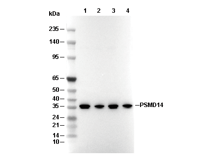 Lane 1: HT1080, Lane 2: LNCAP, Lane 3: RAW264.7, Lane 4: Neuro-2a