|
Comment citer 1. Pour la citation dans le texte (Matériel & Méthodes) : 2. Pour le tableau des ressources clés : |
||
|
Numéro vert : (877) 796-6397 -- États-Unis et Canada uniquement -- |
Fax : +1-832-582-8590 Commandes : +1-832-582-8158 |
Support technique : +1-832-582-8158 Ext:3 Veuillez indiquer votre numéro de commande dans le-mail. Nous nous efforçons de répondre à toutes les demandes par e-mail dans un délai dun jour ouvrable. |
Description biologique
| Spécificité | Phospho-Met (Tyr1349) Antibody [D24L22] détecte les niveaux endogènes de la protéine Met totale uniquement lorsqu'elle est phosphorylée en Tyr1349. |
|---|---|
| Contexte | Phospho-Met (Tyr1349) fait référence à l'état activé du récepteur du facteur de croissance des hépatocytes Met, une récepteur tyrosine kinase hétérodimérique liée par des ponts disulfures (chaîne α extracellulaire d'environ 45 kDa et chaîne β transmembranaire d'environ 145 kDa) qui se lie à HGF pour orchestrer des programmes de croissance invasifs tels que la prolifération, la survie, la migration et la morphogenèse dans les cellules épithéliales et d'autres types de cellules. Met comporte un domaine sémaphorine (SEMA), des répétitions PSI et quatre IPT dans sa région extracellulaire pour la reconnaissance de HGF et la dimérisation, une seule hélice transmembranaire, et un segment intracellulaire avec une région juxtamembranaire (contenant Tyr1003 pour l'ubiquitination/dégradation médiée par CBL et les Ser957/Ser975 inhibitrices), un domaine tyrosine kinase (avec Tyr1234/Tyr1235 de la boucle d'activation dont la phosphorylation améliore l'activité catalytique), et une queue C-terminale abritant Tyr1349 et Tyr1356 qui forment un site de fixation multisubstrat. Lors de la liaison à HGF, Met dimérise et le domaine kinase transphosphoryle Tyr1234/1235 pour stabiliser la conformation active, permettant la phosphorylation ultérieure de Tyr1349 (et Tyr1356). Phospho-Tyr1349 fournit un site de liaison à haute affinité pour l'adaptateur Gab1 et recrute des effecteurs contenant des domaines SH2, y compris GRB2, PI3K p85, SHP2, Src et STAT3, assemblant un hub de signalisation qui connecte Met aux voies Ras-MAPK, PI3K-Akt, STAT et GTPase de la famille Rho pour stimuler la progression du cycle cellulaire, la survie, la motilité et la transition épithélio-mésenchymateuse. La mutation ou la perte de Tyr1349/Tyr1356 altère l'ancrage de Gab1 et l'activation en aval de PI3K/MAPK malgré une activité kinase préservée. |
Informations dutilisation
| Application | WB | Dilution |
|
||
|---|---|---|---|---|---|
| Réactivité | Human, Mouse, Rat | ||||
| Source | Rabbit Monoclonal Antibody | MW | 145 kDa | ||
| Tampon de stockage | PBS, pH 7.2+50% Glycerol+0.05% BSA+0.01% NaN3 | Stockage (À partir de la date de réception) |
-20°C (avoid freeze-thaw cycles), 2 years | ||
| WB |
Experimental Protocol:
Sample preparation
1. Tissue: Lyse the tissue sample by adding an appropriate volume of ice-cold RIPA/NP-40 Lysis Buffer (containing Protease Inhibitor Cocktail, Phosphatase Inhibitor Cocktail),and homogenize the tissue at a low temperature. 2. Adherent cell: Aspirate the culture medium and wash the cells with ice-cold PBS twice. Lyse the cells by adding an appropriate volume of RIPA/NP-40 Lysis Buffer (containing Protease Inhibitor Cocktail, Phosphatase Inhibitor Cocktail) and put the sample on ice for 5 min. 3. Suspension cell: Transfer the culture medium to a pre-cooled centrifuge tube. Centrifuge and aspirate the supernatant. Wash the cells with ice-cold PBS twice. Lyse the cells by adding an appropriate volume of RIPA/NP-40 Lysis Buffer (containing Protease Inhibitor Cocktail, Phosphatase Inhibitor Cocktail) and put the sample on ice for 5 min. 4. Place the lysate into a pre-cooled microcentrifuge tube. Centrifuge at 4°C for 15 min. Collect the supernatant;
5. Remove a small volume of lysate to determine the protein concentration;
6. Combine the lysate with protein loading buffer. Boil 20 µL sample under 95-100°C for 5 min. Centrifuge for 5 min after cool down on ice.
Electrophoretic separation
1. According to the concentration of extracted protein, load appropriate amount of protein sample and marker onto SDS-PAGE gels for electrophoresis. Recommended separating gel (lower gel) concentration: 5%. Reference Table for Selecting SDS-PAGE Separation Gel Concentrations 2. Power up 80V for 30 minutes. Then the power supply is adjusted (110 V~150 V), the Marker is observed, and the electrophoresis can be stopped when the indicator band of the predyed protein Marker where the protein is located is properly separated. (Note that the current should not be too large when electrophoresis, too large current (more than 150 mA) will cause the temperature to rise, affecting the result of running glue. If high currents cannot be avoided, an ice bath can be used to cool the bath.)
Transfer membrane
1. Take out the converter, soak the clip and consumables in the pre-cooled converter;
2. Activate PVDF membrane with methanol for 1 min and rinse with transfer buffer;
3. Install it in the order of "black edge of clip - sponge - filter paper - filter paper - glue -PVDF membrane - filter paper - filter paper - sponge - white edge of clip"; 4. The protein was electrotransferred to PVDF membrane. ( 0.45 µm PVDF membrane is recommended ) Reference Table for Selecting PVDF Membrane Pore Size Specifications Recommended conditions for wet transfer: 200 mA, 120 min. ( Note that the transfer conditions can be adjusted according to the protein size. For high-molecular-weight proteins, a higher current and longer transfer time are recommended. However, ensure that the transfer tank remains at a low temperature to prevent gel melting.)
Block
1. After electrotransfer, wash the film with TBST at room temperature for 5 minutes;
2. Incubate the film in the blocking solution ( recommending 5% BSA solution)
for 1 hour at room temperature;
3. Wash the film with TBST for 3 times, 5 minutes each time.
Antibody incubation
1. Use 5% skim milk powder to prepare the primary antibody working liquid (recommended dilution ratio for primary antibody 1:1000), gently shake and incubate with the film at 4°C overnight; 2. Wash the film with TBST 3 times, 5 minutes each time;
3. Add the secondary antibody to the blocking solution and incubate with the film gently at room temperature for 1 hour;
4. After incubation, wash the film with TBST 3 times for 5 minutes each time.
Antibody staining
1. Add the prepared ECL luminescent substrate (or select other color developing substrate according to the second antibody) and mix evenly;
2. Incubate with the film for 1 minute, remove excess substrate (keep the film moist), wrap with plastic film, and expose in the imaging system.
|
Références
|
Données dapplication
WB
Validé par Selleck
-
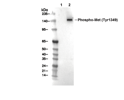 Lane 1: H-4-II-E, Lane 2: H-4-II-E (HGF, 50ng/mL, 5min)