|
Comment citer 1. Pour la citation dans le texte (Matériel & Méthodes) : 2. Pour le tableau des ressources clés : |
||
|
Numéro vert : (877) 796-6397 -- États-Unis et Canada uniquement -- |
Fax : +1-832-582-8590 Commandes : +1-832-582-8158 |
Support technique : +1-832-582-8158 Ext:3 Veuillez indiquer votre numéro de commande dans le-mail. Nous nous efforçons de répondre à toutes les demandes par e-mail dans un délai dun jour ouvrable. |
Description biologique
| Spécificité | p47phox Antibody [L8D20] détecte les niveaux endogènes de la protéine p47phox totale. |
|---|---|
| Contexte | p47phox (facteur cytosolique des neutrophiles 1, NCF1) est une sous-unité organisatrice critique du complexe NADPH-oxydase 2 (NOX2) des phagocytes, essentiel à l'immunité innée en coordonnant l'assemblage des facteurs cytosoliques (p47phox, p67phox, p40phox, Rac) avec le cytochrome b558 lié à la membrane (gp91phox/NOX2-p22phox) pour faciliter la production de superoxyde destiné à l'élimination microbienne ; sa déficience entraîne une granulomatose chronique (GC), caractérisée par des infections récurrentes. La protéine p47phox de 390 acides aminés adopte une conformation auto-inhibée, comprenant un domaine N-terminal d'homologie phox (PX) qui se lie au PI(3,4)P2 ou à l'acide phosphatidique pour le ciblage membranaire, des domaines tandem Src homologie 3 (SH3) qui, à l'état de repos, interagissent avec une région auto-inhibitrice (AIR) pour bloquer l'activité, une région riche en proline C-terminale (PRR) et une queue riche en sérine (Ser303–Ser379) contenant de multiples sites de phosphorylation ainsi que des motifs polybasiques pour l'ancrage initial de p22phox. Lors de la reconnaissance d'un pathogène, une phosphorylation rapide et multi-sites par des kinases telles que PKC, MAPK et PAK sur les sérines C-terminales (notamment Ser303/304/328/359/370) perturbe l'interaction AIR-SH3, expose les domaines SH3 cryptiques pour engager le PRR de p22phox pour la translocation membranaire, libère le domaine PX pour l'ancrage lipidique, et recrute p67phox et Rac pour activer le flavocytochrome pour le transfert d'électrons du NADPH à l'O₂, produisant du superoxyde via une cinétique de ping-pong. Ce commutateur conformationnel, piloté par la phosphorylation, assure le contrôle spatial et temporel de la génération de ROS au niveau des phagosomes, avec des mutations comme Δ219–222 ou W193R dans p47phox qui altèrent la liaison SH3-p22phox et réduisent la production de superoxyde de 60 à 100 %. Les modifications post-traductionnelles, telles que la phosphorylation de Tyr159/240, améliorent encore l'amorçage de l'oxydase. Une activité p47phox dérégulée contribue au stress oxydatif vasculaire dans l'hypertension et l'insuffisance cardiaque par l'activation endothéliale de NOX, favorise l'athérosclérose via les ROS générées par les cellules musculaires lisses vasculaires et les monocytes, et est impliquée dans les maladies inflammatoires où l'hyperactivité de NOX2 entraîne des lésions tissulaires. |
Informations dutilisation
| Application | WB, IP, IHC | Dilution |
|
||||||
|---|---|---|---|---|---|---|---|---|---|
| Réactivité | Human | ||||||||
| Source | Rabbit Monoclonal Antibody | MW | 44 kDa | ||||||
| Tampon de stockage | PBS, pH 7.2+50% Glycerol+0.05% BSA+0.01% NaN3 | Stockage (À partir de la date de réception) |
-20°C (avoid freeze-thaw cycles), 2 years | ||||||
| WB |
Experimental Protocol:
Sample preparation
1. Tissue: Lyse the tissue sample by adding an appropriate volume of ice-cold RIPA/NP-40 Lysis Buffer (containing Protease Inhibitor Cocktail),and homogenize the tissue at a low temperature. 2. Adherent cell: Aspirate the culture medium and wash the cells with ice-cold PBS twice. Lyse the cells by adding an appropriate volume of RIPA/NP-40 Lysis Buffer (containing Protease Inhibitor Cocktail) and put the sample on ice for 5 min. 3. Suspension cell: Transfer the culture medium to a pre-cooled centrifuge tube. Centrifuge and aspirate the supernatant. Wash the cells with ice-cold PBS twice. Lyse the cells by adding an appropriate volume of RIPA/NP-40 Lysis Buffer (containing Protease Inhibitor Cocktail) and put the sample on ice for 5 min. 4. Place the lysate into a pre-cooled microcentrifuge tube. Centrifuge at 4°C for 15 min. Collect the supernatant;
5. Remove a small volume of lysate to determine the protein concentration;
6. Combine the lysate with protein loading buffer. Boil 20 µL sample under 95-100°C for 5 min. Centrifuge for 5 min after cool down on ice.
Electrophoretic separation
1. According to the concentration of extracted protein, load appropriate amount of protein sample and marker onto SDS-PAGE gels for electrophoresis. Recommended separating gel (lower gel) concentration: 10%. Reference Table for Selecting SDS-PAGE Separation Gel Concentrations 2. Power up 80V for 30 minutes. Then the power supply is adjusted (110 V~150 V), the Marker is observed, and the electrophoresis can be stopped when the indicator band of the predyed protein Marker where the protein is located is properly separated. (Note that the current should not be too large when electrophoresis, too large current (more than 150 mA) will cause the temperature to rise, affecting the result of running glue. If high currents cannot be avoided, an ice bath can be used to cool the bath.)
Transfer membrane
1. Take out the converter, soak the clip and consumables in the pre-cooled converter;
2. Activate PVDF membrane with methanol for 1 min and rinse with transfer buffer;
3. Install it in the order of "black edge of clip - sponge - filter paper - filter paper - glue -PVDF membrane - filter paper - filter paper - sponge - white edge of clip"; 4. The protein was electrotransferred to PVDF membrane. ( 0.45 µm PVDF membrane is recommended ) Reference Table for Selecting PVDF Membrane Pore Size Specifications Recommended conditions for wet transfer: 200 mA, 120 min. ( Note that the transfer conditions can be adjusted according to the protein size. For high-molecular-weight proteins, a higher current and longer transfer time are recommended. However, ensure that the transfer tank remains at a low temperature to prevent gel melting.)
Block
1. After electrotransfer, wash the film with TBST at room temperature for 5 minutes;
2. Incubate the film in the blocking solution for 1 hour at room temperature;
3. Wash the film with TBST for 3 times, 5 minutes each time.
Antibody incubation
1. Use 5% skim milk powder to prepare the primary antibody working liquid (recommended dilution ratio for primary antibody 1:1000), gently shake and incubate with the film at 4°C overnight; 2. Wash the film with TBST 3 times, 5 minutes each time;
3. Add the secondary antibody to the blocking solution and incubate with the film gently at room temperature for 1 hour;
4. After incubation, wash the film with TBST 3 times for 5 minutes each time.
Antibody staining
1. Add the prepared ECL luminescent substrate (or select other color developing substrate according to the second antibody) and mix evenly;
2. Incubate with the film for 1 minute, remove excess substrate (keep the film moist), wrap with plastic film, and expose in the imaging system.
|
| IHC |
Experimental Protocol:
Deparaffinization/Rehydration
1. Deparaffinize/hydrate sections:
2. Incubate sections in three washes of xylene for 5 min each.
3. Incubate sections in two washes of 100% ethanol for 10 min each.
4. Incubate sections in two washes of 95% ethanol for 10 min each.
5. Wash sections two times in dH2O for 5 min each.
6.Antigen retrieval: For Citrate: Heat slides in a microwave submersed in 1X citrate unmasking solution until boiling is initiated; continue with 10 min at a sub-boiling temperature (95°-98°C). Cool slides on bench top for 30 min.
Staining
1. Wash sections in dH2O three times for 5 min each.
2. Incubate sections in 3% hydrogen peroxide for 10 min.
3. Wash sections in dH2O two times for 5 min each.
4. Wash sections in wash buffer for 5 min.
5. Block each section with 100–400 µl of blocking solution for 1 hr at room temperature.
6. Remove blocking solution and add 100–400 µl primary antibody diluent in to each section. Incubate overnight at 4°C.
7. Remove antibody solution and wash sections with wash buffer three times for 5 min each.
8. Cover section with 1–3 drops HRPas needed. Incubate in a humidified chamber for 30 min at room temperature.
9. Wash sections three times with wash buffer for 5 min each.
10. Add DAB Chromogen Concentrate to DAB Diluent and mix well before use.
11. Apply 100–400 µl DAB to each section and monitor closely. 1–10 min generally provides an acceptable staining intensity.
12. Immerse slides in dH2O.
13. If desired, counterstain sections with hematoxylin.
14. Wash sections in dH2O two times for 5 min each.
15. Dehydrate sections: Incubate sections in 95% ethanol two times for 10 sec each; Repeat in 100% ethanol, incubating sections two times for 10 sec each; Repeat in xylene, incubating sections two times for 10 sec each.
16. Mount sections with coverslips and mounting medium.
|
Références
|
Données dapplication
WB
Validé par Selleck
-
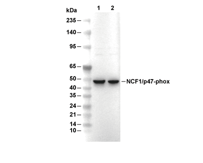 Lane 1: Raji, Lane 2: Ramos