|
Comment citer 1. Pour la citation dans le texte (Matériel & Méthodes) : 2. Pour le tableau des ressources clés : |
||
|
Numéro vert : (877) 796-6397 -- États-Unis et Canada uniquement -- |
Fax : +1-832-582-8590 Commandes : +1-832-582-8158 |
Support technique : +1-832-582-8158 Ext:3 Veuillez indiquer votre numéro de commande dans le-mail. Nous nous efforçons de répondre à toutes les demandes par e-mail dans un délai dun jour ouvrable. |
Description biologique
| Spécificité | JAK3 Antibody [F8D8] détecte les niveaux endogènes de la protéine JAK3 totale. |
|---|---|
| Contexte | JAK3 (Janus kinase 3) est un membre de la famille des Janus kinases (JAK) de tyrosine kinases non-réceptrices, comprenant JAK1, JAK2, JAK3 et TYK2, et est principalement exprimé dans les cellules hématopoïétiques, où il s'associe spécifiquement à la chaîne gamma commune (γc) des récepteurs de cytokines pour l'IL-2, l'IL-4, l'IL-7, l'IL-9, l'IL-15 et l'IL-21. JAK3 présente une organisation multidomaine conservée comprenant un domaine FERM N-terminal pour la liaison au récepteur, un domaine de type SH2, un domaine pseudokinase (JH2) qui manque d'activité catalytique intrinsèque mais régule négativement le domaine kinase actif adjacent (JH1), et un domaine kinase JH1 C-terminal avec une structure bilobée caractérisée par un lobe N en feuillet β, une hélice αC, et un lobe C contenant une hélice αFG insérée (résidus 1029–1038) ; les sites de phosphorylation régulatrice comprennent Tyr980 et Tyr981 dans la boucle d'activation de JH1. La dimérisation du récepteur induite par les cytokines déclenche l'autophosphorylation de JAK3 et la transphosphorylation avec JAK1 ou JAK2, activant le domaine JH1 pour phosphoryler les tyrosines du récepteur, qui servent de sites d'ancrage pour les protéines STAT (telles que STAT5) via les domaines SH2. L'activation subséquente de STAT favorise la transcription génique critique pour la prolifération, la différenciation, la survie et le développement des cellules immunitaires, en particulier dans les lymphocytes T, les lymphocytes B et les cellules NK. Cette voie JAK-STAT est essentielle pour la lymphopoïèse et l'immunité adaptative, avec le domaine pseudokinase JH2 supprimant allostériquement l'activité JH1 jusqu'à l'engagement du ligand. La dérégulation de JAK3, y compris les mutations à gain de fonction (souvent dans JH2), conduit à une signalisation soutenue et à l'oncogenèse dans les leucémies et les lymphomes, tandis que les mutations à perte de fonction entraînent une immunodéficience combinée sévère (SCID). |
Informations dutilisation
| Application | WB, IP | Dilution |
|
||||
|---|---|---|---|---|---|---|---|
| Réactivité | Human | ||||||
| Source | Rabbit Monoclonal Antibody | MW | 115 kDa | ||||
| Tampon de stockage | PBS, pH 7.2+50% Glycerol+0.05% BSA+0.01% NaN3 | Stockage (À partir de la date de réception) |
-20°C (avoid freeze-thaw cycles), 2 years | ||||
| WB |
Experimental Protocol:
Sample preparation
1. Tissue: Lyse the tissue sample by adding an appropriate volume of ice-cold RIPA/NP-40 Lysis Buffer (containing Protease Inhibitor Cocktail),and homogenize the tissue at a low temperature. 2. Adherent cell: Aspirate the culture medium and wash the cells with ice-cold PBS twice. Lyse the cells by adding an appropriate volume of RIPA/NP-40 Lysis Buffer (containing Protease Inhibitor Cocktail) and put the sample on ice for 5 min. 3. Suspension cell: Transfer the culture medium to a pre-cooled centrifuge tube. Centrifuge and aspirate the supernatant. Wash the cells with ice-cold PBS twice. Lyse the cells by adding an appropriate volume of RIPA/NP-40 Lysis Buffer (containing Protease Inhibitor Cocktail) and put the sample on ice for 5 min. 4. Place the lysate into a pre-cooled microcentrifuge tube. Centrifuge at 4°C for 15 min. Collect the supernatant;
5. Remove a small volume of lysate to determine the protein concentration;
6. Combine the lysate with protein loading buffer. Boil 20 µL sample under 95-100°C for 5 min. Centrifuge for 5 min after cool down on ice.
Electrophoretic separation
1. According to the concentration of extracted protein, load appropriate amount of protein sample and marker onto SDS-PAGE gels for electrophoresis. Recommended separating gel (lower gel) concentration: 5%. Reference Table for Selecting SDS-PAGE Separation Gel Concentrations 2. Power up 80V for 30 minutes. Then the power supply is adjusted (110 V~150 V), the Marker is observed, and the electrophoresis can be stopped when the indicator band of the predyed protein Marker where the protein is located is properly separated. (Note that the current should not be too large when electrophoresis, too large current (more than 150 mA) will cause the temperature to rise, affecting the result of running glue. If high currents cannot be avoided, an ice bath can be used to cool the bath.)
Transfer membrane
1. Take out the converter, soak the clip and consumables in the pre-cooled converter;
2. Activate PVDF membrane with methanol for 1 min and rinse with transfer buffer;
3. Install it in the order of "black edge of clip - sponge - filter paper - filter paper - glue -PVDF membrane - filter paper - filter paper - sponge - white edge of clip"; 4. The protein was electrotransferred to PVDF membrane. ( 0.45 µm PVDF membrane is recommended ) Reference Table for Selecting PVDF Membrane Pore Size Specifications Recommended conditions for wet transfer: 200 mA, 120 min. ( Note that the transfer conditions can be adjusted according to the protein size. For high-molecular-weight proteins, a higher current and longer transfer time are recommended. However, ensure that the transfer tank remains at a low temperature to prevent gel melting.)
Block
1. After electrotransfer, wash the film with TBST at room temperature for 5 minutes;
2. Incubate the film in the blocking solution for 1 hour at room temperature;
3. Wash the film with TBST for 3 times, 5 minutes each time.
Antibody incubation
1. Use 5% skim milk powder to prepare the primary antibody working liquid (recommended dilution ratio for primary antibody 1:1000), gently shake and incubate with the film at 4°C overnight; 2. Wash the film with TBST 3 times, 5 minutes each time;
3. Add the secondary antibody to the blocking solution and incubate with the film gently at room temperature for 1 hour;
4. After incubation, wash the film with TBST 3 times for 5 minutes each time.
Antibody staining
1. Add the prepared ECL luminescent substrate (or select other color developing substrate according to the second antibody) and mix evenly;
2. Incubate with the film for 1 minute, remove excess substrate (keep the film moist), wrap with plastic film, and expose in the imaging system.
|
Références
|
Données dapplication
WB
Validé par Selleck
-
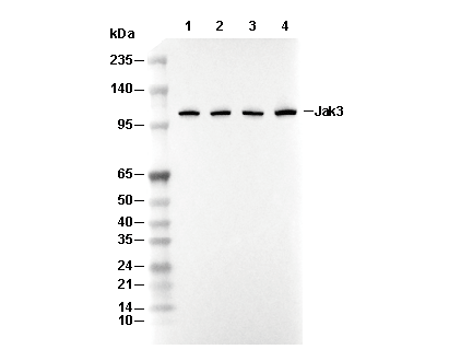 Lane 1: NK-92, Lane 2: Jurkat, Lane 3: SET-2, Lane 4: KARPAS-299