|
Comment citer 1. Pour la citation dans le texte (Matériel & Méthodes) : 2. Pour le tableau des ressources clés : |
||
|
Numéro vert : (877) 796-6397 -- États-Unis et Canada uniquement -- |
Fax : +1-832-582-8590 Commandes : +1-832-582-8158 |
Support technique : +1-832-582-8158 Ext:3 Veuillez indiquer votre numéro de commande dans le-mail. Nous nous efforçons de répondre à toutes les demandes par e-mail dans un délai dun jour ouvrable. |
Description biologique
| Spécificité | ISG15 Antibody [H10C1] détecte les niveaux endogènes de la protéine ISG15 totale. |
|---|---|
| Contexte | Le gène 15 stimulé par l'interféron (ISG15) est une protéine de type ubiquitine qui joue un rôle essentiel dans l'immunité innée et les réponses cellulaires aux infections virales en fonctionnant à la fois comme modificateur intracellulaire par ISGylation et comme cytokine extracellulaire. ISG15 comprend deux domaines de type ubiquitine (Ubl) connectés par une charnière flexible, chaque domaine formant un repliement β-grasp similaire à l'ubiquitine. Les résidus clés dans le domaine C-terminal, tels que Lys90, Trp123 et Phe149, confèrent une spécificité pour la liaison aux enzymes activatrices d'ISG15 comme UbE1L, tandis que le domaine N-terminal exposé au solvant facilite le transfert d'ISG15 aux protéines cibles pendant l'ISGylation, un processus en plusieurs étapes impliquant les ligases E1 (Ube1L), E2 (UbcH8) et E3. L'expression d'ISG15 est fortement induite par les interférons de type I (α/β) et les infections virales. Il peut être sécrété par les cellules immunitaires, où il améliore les réponses immunitaires en stimulant la production d'interféron γ, en favorisant la prolifération des cellules tueuses naturelles et en augmentant la chimiotaxie des neutrophiles. L'ISGylation modifie une variété de protéines, y compris Serpin 2A, PLCγ1, ERK1/2, Jak1 et Stat1, entraînant une activité accrue de Jak1 et Stat1 et une signalisation d'interféron amplifiée, distincte de l'ubiquitination. L'ISGylation ne cible pas les protéines pour la dégradation. La nature réversible de la conjugaison d'ISG15, régulée par la protéase Ubp43, assure un contrôle dynamique des voies de signalisation d'ISG15. |
Informations dutilisation
| Application | WB , IP | Dilution |
|
||||
|---|---|---|---|---|---|---|---|
| Réactivité | Human | ||||||
| Source | Rabbit Monoclonal Antibody | MW | 15 kDa | ||||
| Tampon de stockage | PBS, pH 7.2+50% Glycerol+0.05% BSA+0.01% NaN3 | Stockage (À partir de la date de réception) |
-20°C (avoid freeze-thaw cycles), 2 years | ||||
| WB |
Experimental Protocol:
Sample preparation
1. Tissue: Lyse the tissue sample by adding an appropriate volume of ice-cold RIPA/NP-40 Lysis Buffer (containing Protease Inhibitor Cocktail),and homogenize the tissue at a low temperature. 2. Adherent cell: Aspirate the culture medium and wash the cells with ice-cold PBS twice. Lyse the cells by adding an appropriate volume of RIPA/NP-40 Lysis Buffer (containing Protease Inhibitor Cocktail) and put the sample on ice for 5 min. 3. Suspension cell: Transfer the culture medium to a pre-cooled centrifuge tube. Centrifuge and aspirate the supernatant. Wash the cells with ice-cold PBS twice. Lyse the cells by adding an appropriate volume of RIPA/NP-40 Lysis Buffer (containing Protease Inhibitor Cocktail) and put the sample on ice for 5 min. 4. Place the lysate into a pre-cooled microcentrifuge tube. Centrifuge at 4°C for 15 min. Collect the supernatant;
5. Remove a small volume of lysate to determine the protein concentration;
6. Combine the lysate with protein loading buffer. Boil 20 µL sample under 95-100°C for 5 min. Centrifuge for 5 min after cool down on ice.
Electrophoretic separation
1. According to the concentration of extracted protein, load appropriate amount of protein sample and marker onto SDS-PAGE gels for electrophoresis. Recommended separating gel (lower gel) concentration: 20%. Reference Table for Selecting SDS-PAGE Separation Gel Concentrations 2. Power up 80V for 30 minutes. Then the power supply is adjusted (110 V~150 V), the Marker is observed, and the electrophoresis can be stopped when the indicator band of the predyed protein Marker where the protein is located is properly separated. (Note that the current should not be too large when electrophoresis, too large current (more than 150 mA) will cause the temperature to rise, affecting the result of running glue. If high currents cannot be avoided, an ice bath can be used to cool the bath.)
Transfer membrane
1. Take out the converter, soak the clip and consumables in the pre-cooled converter;
2. Activate PVDF membrane with methanol for 1 min and rinse with transfer buffer;
3. Install it in the order of "black edge of clip - sponge - filter paper - filter paper - glue -PVDF membrane - filter paper - filter paper - sponge - white edge of clip"; 4. The protein was electrotransferred to PVDF membrane. ( 0.22 µm PVDF membrane is recommended )) Reference Table for Selecting PVDF Membrane Pore Size Specifications Recommended conditions for wet transfer: 200 mA, 60 min. ( Note that the transfer conditions can be adjusted according to the protein size. For high-molecular-weight proteins, a higher current and longer transfer time are recommended. However, ensure that the transfer tank remains at a low temperature to prevent gel melting.)
Block
1. After electrotransfer, wash the film with TBST at room temperature for 5 minutes;
2. Incubate the film in the blocking solution for 1 hour at room temperature;
3. Wash the film with TBST for 3 times, 5 minutes each time.
Antibody incubation
1. Use 5% skim milk powder to prepare the primary antibody working liquid (recommended dilution ratio for primary antibody 1:1000), gently shake and incubate with the film at 4°C overnight; 2. Wash the film with TBST 3 times, 5 minutes each time;
3. Add the secondary antibody to the blocking solution and incubate with the film gently at room temperature for 1 hour;
4. After incubation, wash the film with TBST 3 times for 5 minutes each time.
Antibody staining
1. Add the prepared ECL luminescent substrate (or select other color developing substrate according to the second antibody) and mix evenly;
2. Incubate with the film for 1 minute, remove excess substrate (keep the film moist), wrap with plastic film, and expose in the imaging system.
|
Références
|
Données dapplication
WB
Validé par Selleck
-
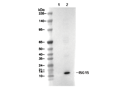 Lane 1: Hela, Lane 2: Hela (hIFNα1, 10 ng/ml, 16 h)