|
Comment citer 1. Pour la citation dans le texte (Matériel & Méthodes) : 2. Pour le tableau des ressources clés : |
||
|
Numéro vert : (877) 796-6397 -- États-Unis et Canada uniquement -- |
Fax : +1-832-582-8590 Commandes : +1-832-582-8158 |
Support technique : +1-832-582-8158 Ext:3 Veuillez indiquer votre numéro de commande dans le-mail. Nous nous efforçons de répondre à toutes les demandes par e-mail dans un délai dun jour ouvrable. |
Description biologique
| Spécificité | Glutamate Receptor 1 (AMPA subtype) Antibody [J12M5] reconnaît les niveaux endogènes de la protéine Glutamate Receptor 1 (AMPA subtype) Rabbit mAb totale. |
|---|---|
| Contexte | Le récepteur du glutamate 1 (GluR1, également connu sous le nom de GluA1) est une sous-unité des récepteurs ionotropes du glutamate de type AMPA (AMPARs), largement exprimés dans les neurones excitateurs, les interneurones et les cellules gliales du système nerveux central. Les AMPARs médient une transmission synaptique excitatrice rapide et sont cruciaux pour la plasticité synaptique, l'apprentissage et la mémoire. Les AMPARs contenant GluA1 peuvent former des assemblages homomériques ou hétéromériques et sont structurellement composés de domaines N-terminaux extracellulaires (NTDs), de domaines de liaison aux ligands (LBDs), de domaines transmembranaires (TMDs) et de domaines C-terminaux intracellulaires. Contrairement aux AMPARs contenant GluA2, les homomères de GluA1 sont perméables au Ca²⁺ et présentent des cinétiques de gating et de désensibilisation distinctes en raison de leurs NTDs très flexibles. GluA1 joue un rôle pivot dans la potentialisation à long terme (LTP) et le ciblage synaptique, avec des sites de phosphorylation clés — tels que Ser818, Ser831, Thr840 et Ser845 — modulant son trafic et son incorporation synaptique de manière dépendante de l'activité. |
Informations dutilisation
| Application | WB, IP, IHC | Dilution |
|
||||||
|---|---|---|---|---|---|---|---|---|---|
| Réactivité | Human, Mouse, Rat | ||||||||
| Source | Rabbit Monoclonal Antibody | MW | 102 kDa | ||||||
| Tampon de stockage | PBS, pH 7.2+50% Glycerol+0.05% BSA+0.01% NaN3 | Stockage (À partir de la date de réception) |
-20°C (avoid freeze-thaw cycles), 2 years | ||||||
| WB |
Experimental Protocol:
Sample preparation
1. Tissue: Lyse the tissue sample by adding an appropriate volume of ice-cold RIPA/NP-40 Lysis Buffer (containing Protease Inhibitor Cocktail),and homogenize the tissue at a low temperature or lyse it by sonication on ice, then incubate on ice for 30 minutes. 2. Adherent cell: Aspirate the culture medium and transfer the cells into an EP tube. Wash the cells with ice-cold PBS twice. Add an appropriate volume of RIPA/NP-40 Lysis Buffer (containing Protease Inhibitor Cocktail), sonicate to lyse the cells, and incubate on ice for 30 minutes. 3. Suspension cell: Transfer the culture medium to a pre-cooled centrifuge tube. Centrifuge and aspirate the supernatant. Wash the cells with ice-cold PBS twice.Add an appropriate volume of RIPA/NP-40 Lysis Buffer (containing Protease Inhibitor Cocktail), sonicate to lyse the cells, and incubate on ice for 30 minutes. 4. Place the lysate into a pre-cooled microcentrifuge tube. Centrifuge at 4°C for 15 min. Collect the supernatant;
5. Remove a small volume of lysate to determine the protein concentration;
6. Combine the lysate with protein loading buffer. Boil 20 µL sample under 95-100°C for 5 min. Centrifuge for 5 min after cool down on ice.
Electrophoretic separation
1. According to the concentration of extracted protein, load appropriate amount of protein sample and marker onto SDS-PAGE gels for electrophoresis. Recommended separating gel (lower gel) concentration: 5%. Reference Table for Selecting SDS-PAGE Separation Gel Concentrations 2. Power up 80V for 30 minutes. Then the power supply is adjusted (110 V~150 V), the Marker is observed, and the electrophoresis can be stopped when the indicator band of the predyed protein Marker where the protein is located is properly separated. (Note that the current should not be too large when electrophoresis, too large current (more than 150 mA) will cause the temperature to rise, affecting the result of running glue. If high currents cannot be avoided, an ice bath can be used to cool the bath.)
Transfer membrane
1. Take out the converter, soak the clip and consumables in the pre-cooled converter;
2. Activate PVDF membrane with methanol for 1 min and rinse with transfer buffer;
3. Install it in the order of "black edge of clip - sponge - filter paper - filter paper - glue -PVDF membrane - filter paper - filter paper - sponge - white edge of clip"; 4. The protein was electrotransferred to PVDF membrane. ( 0.45 µm PVDF membrane is recommended ) Reference Table for Selecting PVDF Membrane Pore Size Specifications Recommended conditions for wet transfer: 200 mA, 120 min. ( Note that the transfer conditions can be adjusted according to the protein size. For high-molecular-weight proteins, a higher current and longer transfer time are recommended. However, ensure that the transfer tank remains at a low temperature to prevent gel melting.)
Block
1. After electrotransfer, wash the film with TBST at room temperature for 5 minutes;
2. Incubate the film in the blocking solution for 1 hour at room temperature;
3. Wash the film with TBST for 3 times, 5 minutes each time.
Antibody incubation
1. Use 5% skim milk powder to prepare the primary antibody working liquid (recommended dilution ratio for primary antibody 1:1000), gently shake and incubate with the film at 4°C overnight; 2. Wash the film with TBST 3 times, 5 minutes each time;
3. Add the secondary antibody to the blocking solution and incubate with the film gently at room temperature for 1 hour;
4. After incubation, wash the film with TBST 3 times for 5 minutes each time.
Antibody staining
1389. Add the prepared ECL luminescent substrate (or select other color developing substrate according to the second antibody) and mix evenly;
2. Incubate with the film for 1 minute, remove excess substrate (keep the film moist), wrap with plastic film, and expose in the imaging system.
|
Références
|
Données dapplication
WB
Validé par Selleck
-
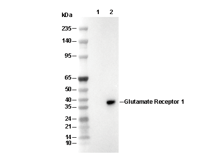 Lane 1: Mouse Glutamate Receptor 2 fragment recombinant protein, Lane 2: Mouse Glutamate Receptor 1 fragment recombinant protein
WB
Validé par Selleck
-
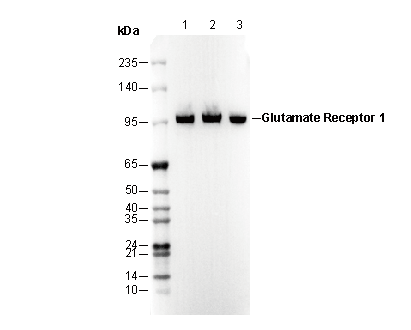 Lane 1: Mouse brain, Lane 2: Rat brain, Lane 3: Human cerebellum