|
Comment citer 1. Pour la citation dans le texte (Matériel & Méthodes) : 2. Pour le tableau des ressources clés : |
||
|
Numéro vert : (877) 796-6397 -- États-Unis et Canada uniquement -- |
Fax : +1-832-582-8590 Commandes : +1-832-582-8158 |
Support technique : +1-832-582-8158 Ext:3 Veuillez indiquer votre numéro de commande dans le-mail. Nous nous efforçons de répondre à toutes les demandes par e-mail dans un délai dun jour ouvrable. |
Description biologique
| Spécificité | CD1c Antibody [H15G15] reconnaît les niveaux endogènes de la protéine CD1c totale. |
|---|---|
| Contexte | CD1c est un membre glycoprotéique de la famille CD1, principalement exprimé sur les cellules présentant l'antigène comme les cellules dendritiques, les macrophages et les cellules B. En tant que protéine transmembranaire de type I, elle comprend une chaîne lourde avec les domaines α1, α2 et α3, ainsi qu'une chaîne légère de β2-microglobuline, formant une fente spécialisée pour la liaison des antigènes lipidiques et glycolipidiques. Contrairement aux molécules MHC classiques présentant des peptides, CD1c présente des antigènes à base de lipides, y compris des composants microbiens (par exemple, le mannosyl-phosphomycokétide mycobactérien) et des auto-lipides. CD1c opère à travers le système endocytaire, capturant les antigènes des compartiments précoces et tardifs via des portails A'/F'/G' uniques dans la fente de liaison, permettant l'accommodation de diverses structures lipidiques. Il active les cellules T restreintes au CD1c, y compris les cellules T γδ et les sous-populations de cellules T αβ, initiant des réponses immunitaires contre les agents pathogènes et les tumeurs. L'asymétrie structurelle gauche-droite dans CD1c permet l'engagement du TCR via un modèle «CD1 altéré», où la liaison lipidique induit des changements conformationnels pour la reconnaissance du TCR. CD1c contribue à la surveillance immunitaire dans les infections, à l'immunité tumorale et au maintien de l'homéostasie tissulaire en modulant les réponses des lymphocytes T auxiliaires, cytotoxiques et régulateurs. Cliniquement, l'expression de CD1c est corrélée avec le pronostic du cancer du sein et l'infiltration immunitaire, tandis que la dérégulation est liée à l'histiocytose et aux infections mycobactériennes. |
Informations dutilisation
| Application | WB, IHC | Dilution |
|
||||
|---|---|---|---|---|---|---|---|
| Réactivité | Human | ||||||
| Source | Rabbit Monoclonal Antibody | MW | 38 kDa | ||||
| Tampon de stockage | PBS, pH 7.2+50% Glycerol+0.05% BSA+0.01% NaN3 | Stockage (À partir de la date de réception) |
-20°C (avoid freeze-thaw cycles), 2 years | ||||
| WB |
Experimental Protocol:
Sample preparation
1. Tissue: Lyse the tissue sample by adding an appropriate volume of ice-cold RIPA/NP-40 Lysis Buffer (containing Protease Inhibitor Cocktail),and homogenize the tissue at a low temperature or lyse it by sonication on ice, then incubate on ice for 30 minutes. 2. Adherent cell: Aspirate the culture medium and transfer the cells into an EP tube. Wash the cells with ice-cold PBS twice. Add an appropriate volume of RIPA/NP-40 Lysis Buffer (containing Protease Inhibitor Cocktail), sonicate to lyse the cells, and incubate on ice for 30 minutes. 3. Suspension cell: Transfer the culture medium to a pre-cooled centrifuge tube. Centrifuge and aspirate the supernatant. Wash the cells with ice-cold PBS twice.Add an appropriate volume of RIPA/NP-40 Lysis Buffer (containing Protease Inhibitor Cocktail), sonicate to lyse the cells, and incubate on ice for 30 minutes. 4. Place the lysate into a pre-cooled microcentrifuge tube. Centrifuge at 4°C for 15 min. Collect the supernatant;
5. Remove a small volume of lysate to determine the protein concentration;
6. Combine the lysate with protein loading buffer. Boil 20 µL sample under 95-100°C for 5 min. Centrifuge for 5 min after cool down on ice.
Electrophoretic separation
1. According to the concentration of extracted protein, load appropriate amount of protein sample and marker onto SDS-PAGE gels for electrophoresis. Recommended separating gel (lower gel) concentration: 10%. Reference Table for Selecting SDS-PAGE Separation Gel Concentrations 2. Power up 80V for 30 minutes. Then the power supply is adjusted (110 V~150 V), the Marker is observed, and the electrophoresis can be stopped when the indicator band of the predyed protein Marker where the protein is located is properly separated. (Note that the current should not be too large when electrophoresis, too large current (more than 150 mA) will cause the temperature to rise, affecting the result of running glue. If high currents cannot be avoided, an ice bath can be used to cool the bath.)
Transfer membrane
1. Take out the converter, soak the clip and consumables in the pre-cooled converter;
2. Activate PVDF membrane with methanol for 1 min and rinse with transfer buffer;
3. Install it in the order of "black edge of clip - sponge - filter paper - filter paper - glue -PVDF membrane - filter paper - filter paper - sponge - white edge of clip"; 4. The protein was electrotransferred to PVDF membrane. ( 0.45 µm PVDF membrane is recommended ) Reference Table for Selecting PVDF Membrane Pore Size Specifications Recommended conditions for wet transfer: 200 mA, 60 min. ( Note that the transfer conditions can be adjusted according to the protein size. For high-molecular-weight proteins, a higher current and longer transfer time are recommended. However, ensure that the transfer tank remains at a low temperature to prevent gel melting.)
Block
1. After electrotransfer, wash the film with TBST at room temperature for 5 minutes;
2. Incubate the film in the blocking solution for 1 hour at room temperature;
3. Wash the film with TBST for 3 times, 5 minutes each time.
Antibody incubation
1. Use 5% skim milk powder to prepare the primary antibody working liquid (recommended dilution ratio for primary antibody 1:1000), gently shake and incubate with the film at 4°C overnight; 2. Wash the film with TBST 3 times, 5 minutes each time;
3. Add the secondary antibody to the blocking solution and incubate with the film gently at room temperature for 1 hour;
4. After incubation, wash the film with TBST 3 times for 5 minutes each time.
Antibody staining
1389. Add the prepared ECL luminescent substrate (or select other color developing substrate according to the second antibody) and mix evenly;
2. Incubate with the film for 1 minute, remove excess substrate (keep the film moist), wrap with plastic film, and expose in the imaging system.
|
Références
|
Données dapplication
WB
Validé par Selleck
-
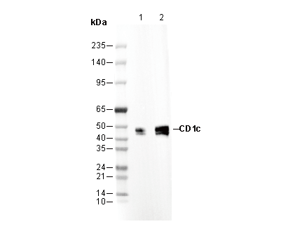 Lane 1: Jurkat, Lane 2: MOLT-4