|
Comment citer 1. Pour la citation dans le texte (Matériel & Méthodes) : 2. Pour le tableau des ressources clés : |
||
|
Numéro vert : (877) 796-6397 -- États-Unis et Canada uniquement -- |
Fax : +1-832-582-8590 Commandes : +1-832-582-8158 |
Support technique : +1-832-582-8158 Ext:3 Veuillez indiquer votre numéro de commande dans le-mail. Nous nous efforçons de répondre à toutes les demandes par e-mail dans un délai dun jour ouvrable. |
Description biologique
| Spécificité | 8-Hydroxy-2'-deoxyguanosine Antibody [K11E8] détecte à la fois la 8-hydroxy-2'-désoxyguanosine (8-OH-dG) endogène et exogène. |
|---|---|
| Contexte | La 8-hydroxy-2'-désoxyguanosine (8-OHdG) est un 2'-désoxyribonucléoside purique modifié formé par l'hydroxylation de la base guanine en position C8 dans l'ADN, résultant typiquement de dommages oxydatifs causés par les espèces réactives de l'oxygène (ROS). Structurellement, elle se compose d'une molécule de désoxyguanosine avec un groupe hydroxyle sur le 8e carbone du cycle de la guanine. La 8-OHdG est largement reconnue comme un biomarqueur clé du stress oxydatif en raison de sa forte prévalence parmi les lésions oxydatives de l'ADN. Elle s'accumule à la fois dans l'ADN nucléaire et mitochondrial et peut être mesurée dans divers échantillons biologiques, y compris l'urine, les tissus et l'ADN des leucocytes. Fonctionnellement, la 8-OHdG sert d'indicateur de dommages endogènes à l'ADN et a été largement utilisée pour évaluer le risque et la progression du cancer, du vieillissement et des maladies dégénératives. Sa présence reflète l'exposition cellulaire aux agents oxydatifs et est essentielle pour évaluer l'impact des facteurs environnementaux et liés au mode de vie sur la stabilité génomique. |
Informations dutilisation
| Application | IHC-P | Dilution |
|
||
|---|---|---|---|---|---|
| Réactivité | Species independent | ||||
| Source | Mouse Monoclonal Antibody | MW | |||
| Tampon de stockage | PBS, pH 7.2+50% Glycerol+0.05% BSA+0.01% NaN3 | Stockage (À partir de la date de réception) |
-20°C (avoid freeze-thaw cycles), 2 years | ||
| IHC |
Experimental Protocol:
Deparaffinization/Rehydration
1. Deparaffinize/hydrate sections:
2. Incubate sections in three washes of xylene for 5 min each.
3. Incubate sections in two washes of 100% ethanol for 10 min each.
4. Incubate sections in two washes of 95% ethanol for 10 min each.
5. Wash sections two times in dH2O for 5 min each.
6.Antigen retrieval: For Citrate: Heat slides in a microwave submersed in 1X citrate unmasking solution until boiling is initiated; continue with 10 min at a sub-boiling temperature (95°-98°C). Cool slides on bench top for 30 min.
Staining
1. Wash sections in dH2O three times for 5 min each.
2. Incubate sections in 3% hydrogen peroxide for 10 min.
3. Wash sections in dH2O two times for 5 min each.
4. Wash sections in wash buffer for 5 min.
5. Block each section with 100–400 µl of blocking solution for 1 hr at room temperature.
6. Remove blocking solution and add 100–400 µl primary antibody diluent in to each section. Incubate overnight at 4°C.
7. Remove antibody solution and wash sections with wash buffer three times for 5 min each.
8. Cover section with 1–3 drops HRPas needed. Incubate in a humidified chamber for 30 min at room temperature.
9. Wash sections three times with wash buffer for 5 min each.
10. Add DAB Chromogen Concentrate to DAB Diluent and mix well before use.
11. Apply 100–400 µl DAB to each section and monitor closely. 1–10 min generally provides an acceptable staining intensity.
12. Immerse slides in dH2O.
13. If desired, counterstain sections with hematoxylin.
14. Wash sections in dH2O two times for 5 min each.
15. Dehydrate sections: Incubate sections in 95% ethanol two times for 10 sec each; Repeat in 100% ethanol, incubating sections two times for 10 sec each; Repeat in xylene, incubating sections two times for 10 sec each.
16. Mount sections with coverslips and mounting medium.
|
Références
|
Données dapplication
IHC
Validé par Selleck
-
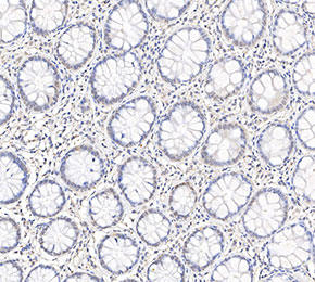 Immunohistochemical analysis of formalin fixed paraffin embedded human colorectal cancer tissue with F1714 at 1:50 dilution.