|
Hoe te citeren 1. Voor in-tekst citatie (materialen en methoden): 2. Voor de tabel met belangrijke bronnen: |
||
|
Gratis nummer: (877) 796-6397 -- Alleen VS en Canada -- |
Fax: +1-832-582-8590 Bestellingen: +1-832-582-8158 |
Technische ondersteuning: +1-832-582-8158 Ext:3 Gelieve uw bestelnummer in de e-mail te vermelden. Wij streven ernaar alle e-mailvragen binnen één werkdag te beantwoorden. |
Biologische Beschrijving
| Specificiteit | YTHDF1 Antibody [H2N8] detecteert totale endogene niveaus van YTHDF1-eiwit. |
|---|---|
| Achtergrond | YTHDF1 is een goed bestudeerd m6A-reader-eiwit dat cruciaal is voor eiwitvertaling, zelfvernieuwing van stamcellen en embryonale ontwikkeling. Het reguleert de expressie van doelgenen via meerdere mechanismen, waaronder het verbeteren van eiwitvertaling en het moduleren van mRNA-stabiliteit. De cellulaire abundantie van YTHDF1 wordt strak gecontroleerd door ingewikkelde netwerken die transcriptionele, post-transcriptionele en post-translationele regulatie omvatten. YTHDF1 is de meest voorkomende m6A-reader, die m6A-gemodificeerd mRNA verbindt met zijn stroomafwaartse bestemming, met name bij eiwitvertaling. Het vergemakkelijkt de vorming van het eIF3b-translatie-initiatiecomplex, waardoor de translatie van belangrijke doelen zoals YAP wordt bevorderd. Dit proces kan worden verstoord door eIF3b uit te putten, YTHDF1 te remmen of METTL3-gemedieerde translatiegebeurtenissen te verstoren. Bovendien speelt YTHDF1 een vitale rol in de RNA-stabiliteit, aangezien het de stabiliteit en expressie van c-Myc mRNA verhoogt, een effect gedreven door METTL3-gekatalyseerde m6A-modificatie. Andere YTH-familie-eiwitten vertonen ook verschillende rollen, zoals YTHDC1 dat voornamelijk functioneert bij RNA-splicing en nucleair export, met lokalisatie voornamelijk in de nucleus, terwijl YTHDC2 essentieel is voor m6A-gemodificeerd mRNA-translatie en -afbraak, met name binnen verwerkingslichamen, en YTHDF3 werkt samen met YTHDF1 om mRNA-translatie te verbeteren en vergemakkelijkt de afbraak van m6A-gemodificeerde transcripten via YTHDF2. Daarom onderstrepen deze eiwitten de veelzijdige rollen van m6A-lezers in RNA-regulatie, waarbij YTHDF1 een belangrijke speler is in embryonale ontwikkeling, stamcelonderhoud en kankerprogressie. |
Gebruiksinformatie
| Toepassing | WB, IP | Verdunning |
|
||||
|---|---|---|---|---|---|---|---|
| Reactiviteit | Human, Mouse, Rat | ||||||
| Bron | Rabbit Monoclonal Antibody | MW | 61 kDa | ||||
| Opslagbuffer | PBS, pH 7.2+50% Glycerol+0.05% BSA+0.01% NaN₃ | Opslag (Vanaf de datum van ontvangst) |
-20°C (avoid freeze-thaw cycles), 2 years | ||||
| WB |
Experimental Protocol:
Sample preparation
1. Tissue: Lyse the tissue sample by adding an appropriate volume of ice-cold RIPA/Tris-HCL Lysis Buffer (containing Protease Inhibitor Cocktail),and homogenize the tissue at a low temperature or lyse it by sonication on ice, then incubate on ice for 30 minutes. 2. Adherent cell: Aspirate the culture medium and transfer the cells into an EP tube. Wash the cells with ice-cold PBS twice. Add an appropriate volume of RIPA/Tris-HCL Lysis Buffer (containing Protease Inhibitor Cocktail), sonicate to lyse the cells, and incubate on ice for 30 minutes. 3. Suspension cell: Transfer the culture medium to a pre-cooled centrifuge tube. Centrifuge and aspirate the supernatant. Wash the cells with ice-cold PBS twice.Add an appropriate volume of RIPA/Tris-HCL Lysis Buffer (containing Protease Inhibitor Cocktail), sonicate to lyse the cells, and incubate on ice for 30 minutes. 4. Place the lysate into a pre-cooled microcentrifuge tube. Centrifuge at 4°C for 15 min. Collect the supernatant;
5. Remove a small volume of lysate to determine the protein concentration;
6. Combine the lysate with protein loading buffer. Boil 20 µL sample under 95-100°C for 5 min. Centrifuge for 5 min after cool down on ice.
Electrophoretic separation
1. According to the concentration of extracted protein, load appropriate amount of protein sample and marker onto SDS-PAGE gels for electrophoresis. Recommended separating gel (lower gel) concentration: 10%. Reference Table for Selecting SDS-PAGE Separation Gel Concentrations 2. Power up 80V for 30 minutes. Then the power supply is adjusted (110 V~150 V), the Marker is observed, and the electrophoresis can be stopped when the indicator band of the predyed protein Marker where the protein is located is properly separated. (Note that the current should not be too large when electrophoresis, too large current (more than 150 mA) will cause the temperature to rise, affecting the result of running glue. If high currents cannot be avoided, an ice bath can be used to cool the bath.)
Transfer membrane
1. Take out the converter, soak the clip and consumables in the pre-cooled converter;
2. Activate PVDF membrane with methanol for 1 min and rinse with transfer buffer;
3. Install it in the order of "black edge of clip - sponge - filter paper - filter paper - glue -PVDF membrane - filter paper - filter paper - sponge - white edge of clip"; 4. The protein was electrotransferred to PVDF membrane. ( 0.45 µm PVDF membrane is recommended ) Reference Table for Selecting PVDF Membrane Pore Size Specifications Recommended conditions for wet transfer: 200 mA, 120 min. ( Note that the transfer conditions can be adjusted according to the protein size. For high-molecular-weight proteins, a higher current and longer transfer time are recommended. However, ensure that the transfer tank remains at a low temperature to prevent gel melting.)
Block
1. After electrotransfer, wash the film with TBST at room temperature for 5 minutes;
2. Incubate the film in the blocking solution for 1 hour at room temperature;
3. Wash the film with TBST for 3 times, 5 minutes each time.
Antibody incubation
1. Use 5% skim milk powder to prepare the primary antibody working liquid (recommended dilution ratio for primary antibody 1:1000), gently shake and incubate with the film at 4°C overnight; 2. Wash the film with TBST 3 times, 5 minutes each time;
3. Add the secondary antibody to the blocking solution and incubate with the film gently at room temperature for 1 hour;
4. After incubation, wash the film with TBST 3 times for 5 minutes each time.
Antibody staining
1234. Add the prepared ECL luminescent substrate (or select other color developing substrate according to the second antibody) and mix evenly;
2. Incubate with the film for 1 minute, remove excess substrate (keep the film moist), wrap with plastic film, and expose in the imaging system.
|
Referenties
|
Toepassingsgegevens
WB
Gevalideerd door Selleck
-
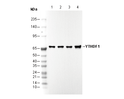 Lane 1: HeLa
Lane 2: 293T
Lane 3: NIH/3T3
Lane 4: PC-12