|
Hoe te citeren 1. Voor in-tekst citatie (materialen en methoden): 2. Voor de tabel met belangrijke bronnen: |
||
|
Gratis nummer: (877) 796-6397 -- Alleen VS en Canada -- |
Fax: +1-832-582-8590 Bestellingen: +1-832-582-8158 |
Technische ondersteuning: +1-832-582-8158 Ext:3 Gelieve uw bestelnummer in de e-mail te vermelden. Wij streven ernaar alle e-mailvragen binnen één werkdag te beantwoorden. |
Biologische Beschrijving
| Specificiteit | VE-cadherin Antibody [C2J1] herkent endogene niveaus van totaal VE-cadherin eiwit. |
|---|---|
| Achtergrond | VE-cadherin, ook bekend als vasculair endotheel cadherin of CD144, is een cruciaal transmembraanproteïne dat voornamelijk tot expressie komt in endotheelcellen, waar het een vitale rol speelt bij het handhaven van cel-celverbindingen die de vasculaire permeabiliteit en integriteit reguleren. Als lid van de cadherine superfamilie heeft VE-cadherin een unieke structuur die bestaat uit een extracellulair domein dat verantwoordelijk is voor homofiele binding aan aangrenzende VE-cadherin moleculen, een enkel transmembraandomein en een intracellulair domein dat interageert met catenines zoals β-catenine en p120-catenine. Deze interactie verankert VE-cadherin aan het actine cytoskelet, wat sterke adhesie tussen endotheelcellen vergemakkelijkt en adherens juncties vormt die essentieel zijn voor de stabiliteit van de endotheelbarrière. VE-cadherin is cruciaal voor angiogenese door de rijping en stabilisatie van vasculaire structuren te reguleren. Het speelt ook een belangrijke rol bij ontsteking door het moduleren van leukocyten-trafficking over het endotheel; tijdens ontstekingsreacties kan fosforylering van VE-cadherin de cel-celadhesie verzwakken, waardoor immuuncellen kunnen transmigreren naar plaatsen van letsel of infectie. De regulatie van VE-cadherin activiteit omvat meerdere mechanismen, waaronder fosforylering door kinasen zoals Src, wat de adhesieve eigenschappen en permeabiliteit beïnvloedt als reactie op groeifactoren zoals VEGF. Dysregulatie van VE-cadherin resulteert in kanker en ontstekingsziekten. |
Gebruiksinformatie
| Toepassing | WB, IP, FCM, ELISA | Verdunning |
|
||||||
|---|---|---|---|---|---|---|---|---|---|
| Reactiviteit | Human, Mouse, Rat, Porcine | ||||||||
| Bron | Mouse Monoclonal Antibody | MW | 130 kDa | ||||||
| Opslagbuffer | PBS, pH 7.2+50% Glycerol+0.05% BSA+0.01% NaN₃ | Opslag (Vanaf de datum van ontvangst) |
-20°C (avoid freeze-thaw cycles), 2 years | ||||||
| WB |
Experimental Protocol:
Sample preparation
1. Tissue: Lyse the tissue sample by adding an appropriate volume of ice-cold RIPA/NP-40 Lysis Buffer (containing Protease Inhibitor Cocktail),and homogenize the tissue at a low temperature or lyse it by sonication on ice, then incubate on ice for 30 minutes. 2. Adherent cell: Aspirate the culture medium and transfer the cells into an EP tube. Wash the cells with ice-cold PBS twice. Add an appropriate volume of RIPA/NP-40 Lysis Buffer (containing Protease Inhibitor Cocktail), sonicate to lyse the cells, and incubate on ice for 30 minutes. 3. Suspension cell: Transfer the culture medium to a pre-cooled centrifuge tube. Centrifuge and aspirate the supernatant. Wash the cells with ice-cold PBS twice.Add an appropriate volume of RIPA/NP-40 Lysis Buffer (containing Protease Inhibitor Cocktail), sonicate to lyse the cells, and incubate on ice for 30 minutes. 4. Place the lysate into a pre-cooled microcentrifuge tube. Centrifuge at 4°C for 15 min. Collect the supernatant;
5. Remove a small volume of lysate to determine the protein concentration;
6. Combine the lysate with protein loading buffer. Boil 20 µL sample under 95-100°C for 5 min. Centrifuge for 5 min after cool down on ice.
Electrophoretic separation
1. According to the concentration of extracted protein, load appropriate amount of protein sample and marker onto SDS-PAGE gels for electrophoresis. Recommended separating gel (lower gel) concentration: 5%. Reference Table for Selecting SDS-PAGE Separation Gel Concentrations 2. Power up 80V for 30 minutes. Then the power supply is adjusted (110 V~150 V), the Marker is observed, and the electrophoresis can be stopped when the indicator band of the predyed protein Marker where the protein is located is properly separated. (Note that the current should not be too large when electrophoresis, too large current (more than 150 mA) will cause the temperature to rise, affecting the result of running glue. If high currents cannot be avoided, an ice bath can be used to cool the bath.)
Transfer membrane
1. Take out the converter, soak the clip and consumables in the pre-cooled converter;
2. Activate PVDF membrane with methanol for 1 min and rinse with transfer buffer;
3. Install it in the order of "black edge of clip - sponge - filter paper - filter paper - glue -PVDF membrane - filter paper - filter paper - sponge - white edge of clip"; 4. The protein was electrotransferred to PVDF membrane. ( 0.45 µm PVDF membrane is recommended ) Reference Table for Selecting PVDF Membrane Pore Size Specifications Recommended conditions for wet transfer: 200 mA, 120 min. ( Note that the transfer conditions can be adjusted according to the protein size. For high-molecular-weight proteins, a higher current and longer transfer time are recommended. However, ensure that the transfer tank remains at a low temperature to prevent gel melting.)
Block
1. After electrotransfer, wash the film with TBST at room temperature for 5 minutes;
2. Incubate the film in the blocking solution for 1 hour at room temperature;
3. Wash the film with TBST for 3 times, 5 minutes each time.
Antibody incubation
1. Use 5% skim milk powder to prepare the primary antibody working liquid (recommended dilution ratio for primary antibody 1:1000), gently shake and incubate with the film at 4°C overnight; 2. Wash the film with TBST 3 times, 5 minutes each time;
3. Add the secondary antibody to the blocking solution and incubate with the film gently at room temperature for 1 hour;
4. After incubation, wash the film with TBST 3 times for 5 minutes each time.
Antibody staining
1387. Add the prepared ECL luminescent substrate (or select other color developing substrate according to the second antibody) and mix evenly;
2. Incubate with the film for 1 minute, remove excess substrate (keep the film moist), wrap with plastic film, and expose in the imaging system. (Exposure time of at least 60s is recommended)
|
Referenties
|
Toepassingsgegevens
WB
Gevalideerd door Selleck
-
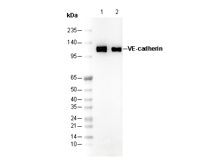 Lane 1: HUV-EC-C
Lane 2: Human placenta