|
Hoe te citeren 1. Voor in-tekst citatie (materialen en methoden): 2. Voor de tabel met belangrijke bronnen: |
||
|
Gratis nummer: (877) 796-6397 -- Alleen VS en Canada -- |
Fax: +1-832-582-8590 Bestellingen: +1-832-582-8158 |
Technische ondersteuning: +1-832-582-8158 Ext:3 Gelieve uw bestelnummer in de e-mail te vermelden. Wij streven ernaar alle e-mailvragen binnen één werkdag te beantwoorden. |
Biologische Beschrijving
| Specificiteit | TSH Receptor/TSH-R Antibody [L10M22] detecteert endogene niveaus van totaal TSH Receptor/TSH-R eiwit. |
|---|---|
| Achtergrond | De schildklierstimulerend hormoonreceptor (TSHR) is een belangrijke regulator van het schildklierhormoonmetabolisme en dient als primaire controller van de schildkliercelfunctie en -groei. Het behoort tot de familie van G-proteïne-gekoppelde receptoren met zeven transmembraandomeinen en is gepositioneerd op het basolaterale membraan van schildklierfollikelcellen. Het TSHR-eiwit is samengesteld uit 764 aminozuren, heeft een molecuulgewicht van ongeveer 87 kDa en medieert zijn effecten via interactie met meerdere G-proteïne-subtypen, met name Gαs en Gαq. Bij activering door TSH initieert de receptor intracellulaire signalering via deze G-proteïnen, waardoor de activiteit van stroomafwaartse effectormoleculen wordt gemoduleerd. De Gαs-route stimuleert de cyclisch adenosinemonofosfaat (cAMP)-cascade, terwijl de Gαq-route de fosfolipase C (PLC)-cascade activeert. Bij verhoogde TSH-concentraties bindt cAMP aan proteïnekinase A (PKA), dat verschillende doeleffectoren fosforyleert, waardoor hun katalytische activiteit wordt verbeterd. Parallel daaraan genereert PLC-activering inositol 1,4,5-trifosfaat (IP₃) en diacylglycerol (DAG), waardoor cellulaire reacties verder worden versterkt. TSHR-expressie wordt positief gereguleerd door fysiologische TSH-niveaus, maar wordt neerwaarts gereguleerd bij aanhoudend hoge TSH-concentraties. Chronische overstimulatie van TSHR, met name via de cAMP-route, kan leiden tot overmatige schildklierhormoonsecretie, schildklierfollikelhyperplasie en klinische hyperthyreoïdie. Mutaties in het TSHR-gen kunnen de eiwitstructuur van de receptor of de post-translationele modificaties ervan beïnvloeden, waardoor de receptorfunctie verandert. Hoewel TSHR niet direct carcinogenese initieert, kan het aanzienlijk bijdragen aan tumorgroei wanneer oncogenen al zijn geactiveerd. |
Gebruiksinformatie
| Toepassing | IHC | Verdunning |
|
||
|---|---|---|---|---|---|
| Reactiviteit | Human | ||||
| Bron | Rabbit Monoclonal Antibody | MW | |||
| Opslagbuffer | PBS, pH 7.2+50% Glycerol+0.05% BSA+0.01% NaN3 | Opslag (Vanaf de datum van ontvangst) |
-20°C (avoid freeze-thaw cycles), 2 years | ||
| IHC |
Experimental Protocol:
Deparaffinization/Rehydration
1. Deparaffinize/hydrate sections:
2. Incubate sections in three washes of xylene for 5 min each.
3. Incubate sections in two washes of 100% ethanol for 10 min each.
4. Incubate sections in two washes of 95% ethanol for 10 min each.
5. Wash sections two times in dH2O for 5 min each.
6.Antigen retrieval: For Citrate: Heat slides in a microwave submersed in 1X citrate unmasking solution until boiling is initiated; continue with 10 min at a sub-boiling temperature (95°-98°C). Cool slides on bench top for 30 min.
Staining
1. Wash sections in dH2O three times for 5 min each.
2. Incubate sections in 3% hydrogen peroxide for 10 min.
3. Wash sections in dH2O two times for 5 min each.
4. Wash sections in wash buffer for 5 min.
5. Block each section with 100–400 µl of blocking solution for 1 hr at room temperature.
6. Remove blocking solution and add 100–400 µl primary antibody diluent in to each section. Incubate overnight at 4°C.
7. Remove antibody solution and wash sections with wash buffer three times for 5 min each.
8. Cover section with 1–3 drops HRPas needed. Incubate in a humidified chamber for 30 min at room temperature.
9. Wash sections three times with wash buffer for 5 min each.
10. Add DAB Chromogen Concentrate to DAB Diluent and mix well before use.
11. Apply 100–400 µl DAB to each section and monitor closely. 1–10 min generally provides an acceptable staining intensity.
12. Immerse slides in dH2O.
13. If desired, counterstain sections with hematoxylin.
14. Wash sections in dH2O two times for 5 min each.
15. Dehydrate sections: Incubate sections in 95% ethanol two times for 10 sec each; Repeat in 100% ethanol, incubating sections two times for 10 sec each; Repeat in xylene, incubating sections two times for 10 sec each.
16. Mount sections with coverslips and mounting medium.
|
Referenties
|
Toepassingsgegevens
IHC
Gevalideerd door Selleck
-
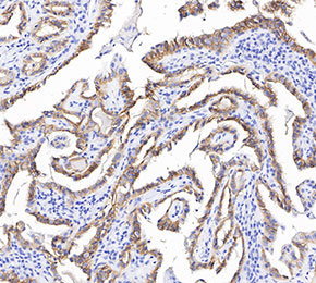 Immunohistochemical analysis of formalin fixed paraffin embedded human thyroid gland tissue with F3696 at 1:1000 dilution.