|
Hoe te citeren 1. Voor in-tekst citatie (materialen en methoden): 2. Voor de tabel met belangrijke bronnen: |
||
|
Gratis nummer: (877) 796-6397 -- Alleen VS en Canada -- |
Fax: +1-832-582-8590 Bestellingen: +1-832-582-8158 |
Technische ondersteuning: +1-832-582-8158 Ext:3 Gelieve uw bestelnummer in de e-mail te vermelden. Wij streven ernaar alle e-mailvragen binnen één werkdag te beantwoorden. |
Biologische Beschrijving
| Specificiteit | Phospho-TFEB (Ser211) Antibody [N21G19] herkent endogene niveaus van TFEB-eiwit alleen wanneer gefosforyleerd op Ser211. |
|---|---|
| Achtergrond | Phospho-TFEB (Ser211) verwijst naar de gefosforyleerde vorm van transcriptiefactor EB (TFEB) op serine-residu 211, een kritieke regulerende plek binnen de structuur ervan. TFEB is een basische helix-loop-helix-leucine zipper (bHLH-Zip) transcriptiefactor van de MiT/TFE-familie die genen reguleert die betrokken zijn bij Autophagy, lysosomale biogenese en cellulaire klaring. Het volledige TFEB-eiwit bevat DNA-bindings-, dimerisatie- en transactivatiedomeinen, waarbij Ser211 zich bevindt in een regio die de subcellulaire lokalisatie reguleert. Structureel gedefinieerd als een Gln glutamine-rijke regio, AD zure domein, bHLH basische helix-loop-helix, LZ leucine zipper domein, Pro proline-rijke segment. Onder voedselrijke omstandigheden fosforyleert mechanistisch target van rapamycine complex 1 (mTORC1) Ser211 op het lysosomale oppervlak, waardoor een bindingsplaats ontstaat voor 14-3-3 chaperonne-eiwitten. Deze interactie maskeert het nucleaire lokalisatiesignaal (NLS) van TFEB, sequestert het in het cytoplasma en voorkomt transcriptionele activering van zijn doelgenen. Wanneer cellen stress of honger ondergaan, neemt de mTORC1-activiteit af, wat leidt tot Ser211-defosforylering, vrijlating van 14-3-3 en nucleaire translocatie van TFEB. Daar activeert TFEB genen die het CLEAR-motief bevatten, waardoor Autophagy en lysosomale functie worden verbeterd. Aldus is fosforylering van Ser211 essentieel voor het dynamisch reguleren van de activiteit van TFEB als reactie op cellulaire metabolische toestanden, het handhaven van energiehomeostase en het vergemakkelijken van de klaring van toxische substraten bij ziekten zoals neurodegeneratie en nieraandoeningen. |
Gebruiksinformatie
| Toepassing | WB | Verdunning |
|
||
|---|---|---|---|---|---|
| Reactiviteit | Human | ||||
| Bron | Rabbit Monoclonal Antibody | MW | 70 kDa | ||
| Opslagbuffer | PBS, pH 7.2+50% Glycerol+0.05% BSA+0.01% NaN3 | Opslag (Vanaf de datum van ontvangst) |
-20°C (avoid freeze-thaw cycles), 2 years | ||
| WB |
Experimental Protocol:
Sample preparation
1. Tissue: Lyse the tissue sample by adding an appropriate volume of ice-cold RIPA/NP-40 Lysis Buffer (containing Protease Inhibitor Cocktail, Phosphatase Inhibitor Cocktail),and homogenize the tissue at a low temperature. 2. Adherent cell: Aspirate the culture medium and wash the cells with ice-cold PBS twice. Lyse the cells by adding an appropriate volume of RIPA/NP-40 Lysis Buffer (containing Protease Inhibitor Cocktail, Phosphatase Inhibitor Cocktail) and put the sample on ice for 5 min. 3. Suspension cell: Transfer the culture medium to a pre-cooled centrifuge tube. Centrifuge and aspirate the supernatant. Wash the cells with ice-cold PBS twice. Lyse the cells by adding an appropriate volume of RIPA/NP-40 Lysis Buffer (containing Protease Inhibitor Cocktail, Phosphatase Inhibitor Cocktail) and put the sample on ice for 5 min. 4. Place the lysate into a pre-cooled microcentrifuge tube. Centrifuge at 4°C for 15 min. Collect the supernatant;
5. Remove a small volume of lysate to determine the protein concentration;
6. Combine the lysate with protein loading buffer. Boil 20 µL sample under 95-100°C for 5 min. Centrifuge for 5 min after cool down on ice.
Electrophoretic separation
1. According to the concentration of extracted protein, load appropriate amount of protein sample and marker onto SDS-PAGE gels for electrophoresis. Recommended separating gel (lower gel) concentration: 10%. Reference Table for Selecting SDS-PAGE Separation Gel Concentrations 2. Power up 80V for 30 minutes. Then the power supply is adjusted (110 V~150 V), the Marker is observed, and the electrophoresis can be stopped when the indicator band of the predyed protein Marker where the protein is located is properly separated. (Note that the current should not be too large when electrophoresis, too large current (more than 150 mA) will cause the temperature to rise, affecting the result of running glue. If high currents cannot be avoided, an ice bath can be used to cool the bath.)
Transfer membrane
1. Take out the converter, soak the clip and consumables in the pre-cooled converter;
2. Activate PVDF membrane with methanol for 1 min and rinse with transfer buffer;
3. Install it in the order of "black edge of clip - sponge - filter paper - filter paper - glue -PVDF membrane - filter paper - filter paper - sponge - white edge of clip"; 4. The protein was electrotransferred to PVDF membrane. ( 0.45 µm PVDF membrane is recommended ) Reference Table for Selecting PVDF Membrane Pore Size Specifications Recommended conditions for wet transfer: 200 mA, 120 min. ( Note that the transfer conditions can be adjusted according to the protein size. For high-molecular-weight proteins, a higher current and longer transfer time are recommended. However, ensure that the transfer tank remains at a low temperature to prevent gel melting.)
Block
1. After electrotransfer, wash the film with TBST at room temperature for 5 minutes;
2. Incubate the film in the blocking solution ( recommending 5% BSA solution)
for 1 hour at room temperature;
3. Wash the film with TBST for 3 times, 5 minutes each time.
Antibody incubation
1. Use 5% skim milk powder to prepare the primary antibody working liquid (recommended dilution ratio for primary antibody 1:1000), gently shake and incubate with the film at 4°C overnight; 2. Wash the film with TBST 3 times, 5 minutes each time;
3. Add the secondary antibody to the blocking solution and incubate with the film gently at room temperature for 1 hour;
4. After incubation, wash the film with TBST 3 times for 5 minutes each time.
Antibody staining
1. Add the prepared ECL luminescent substrate (or select other color developing substrate according to the second antibody) and mix evenly;
2. Incubate with the film for 1 minute, remove excess substrate (keep the film moist), wrap with plastic film, and expose in the imaging system.
|
Referenties
|
Toepassingsgegevens
WB
Gevalideerd door Selleck
-
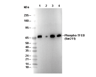 Lane 1: Raji, Lane 2: Raji (Torin 1, 250 nM, 5 h), Lane 3: Daudi, Lane 4: Daudi (Torin 1, 250 nM, 5 h)