|
Hoe te citeren 1. Voor in-tekst citatie (materialen en methoden): 2. Voor de tabel met belangrijke bronnen: |
||
|
Gratis nummer: (877) 796-6397 -- Alleen VS en Canada -- |
Fax: +1-832-582-8590 Bestellingen: +1-832-582-8158 |
Technische ondersteuning: +1-832-582-8158 Ext:3 Gelieve uw bestelnummer in de e-mail te vermelden. Wij streven ernaar alle e-mailvragen binnen één werkdag te beantwoorden. |
Biologische Beschrijving
| Specificiteit | Phospho-CDC37 (Ser13) Antibody [G22K18] detecteert endogene niveaus van CDC37-eiwit alleen wanneer gefosforyleerd op Ser13. |
|---|---|
| Achtergrond | Heat shock proteïne 90 (Hsp90) is een centraal moleculair chaperone dat de vouwing, rijping en stabiliteit van talrijke cliëntproteïnen, met name signaalmoleculen en kinasen, regelt. De activiteit ervan wordt strak gereguleerd door een netwerk van co-chaperones die de rekrutering van cliënten en conformationele overgangen coördineren. Hiertussen vertegenwoordigt celdivisiecyclus 37 (Cdc37, ook bekend als p50) een hooggespecialiseerd co-chaperone met een dominante rol in de regulatie van proteïnekinasen. Als kinase-specifieke adapter rekruteert Cdc37 een breed spectrum aan kinasen naar het Hsp90-complex, waardoor ze worden beschermd tegen afbraak en hun stabilisatie en functionele rijping mogelijk worden gemaakt. Deze rekrutering wordt als onmisbaar beschouwd, aangezien de meeste kinasen afhankelijk zijn van het Cdc37–Hsp90-complex voor een juiste vouwing en activering. Gecodeerd door het CDC37-gen, is Cdc37 functioneel gekoppeld aan celproliferatie en -overleving. In gist is het vereist voor de progressie van de Cell Cycle door belangrijke kinasecomplexen te stabiliseren die essentieel zijn voor mitotische regulatie. In zoogdiersystemen moduleert het de ATPase-activiteit van Hsp90, waardoor het conformationele veranderingen beïnvloedt die cruciaal zijn voor een efficiënte kinasebelasting. Verlies of onderdrukking van CDC37-expressie resulteert in defecte kinasesignalering, groeistop en apoptose. Omgekeerd bevordert verhoogde expressie celproliferatie en is het in verband gebracht met oncogene processen, met name bij prostaatkanker, waar opregulatie van Cdc37 hyperplasie en dysplasie veroorzaakt. Zo fungeert Cdc37 als een cruciale mediator van Hsp90-afhankelijke kinaseregulatie, waarbij moleculaire chaperone-activiteit wordt geïntegreerd met Cell Cycle-controle en tumorigenese. |
Gebruiksinformatie
| Toepassing | WB | Verdunning |
|
||
|---|---|---|---|---|---|
| Reactiviteit | Human, Mouse, Rat | ||||
| Bron | Rabbit Monoclonal Antibody | MW | 50 kDa | ||
| Opslagbuffer | PBS, pH 7.2+50% Glycerol+0.05% BSA+0.01% NaN3 | Opslag (Vanaf de datum van ontvangst) |
-20°C (avoid freeze-thaw cycles), 2 years | ||
| WB |
Experimental Protocol:
Sample preparation
1. Tissue: Lyse the tissue sample by adding an appropriate volume of ice-cold RIPA/NP-40 Lysis Buffer (containing Protease Inhibitor Cocktail, Phosphatase Inhibitor Cocktail),and homogenize the tissue at a low temperature. 2. Adherent cell: Aspirate the culture medium and wash the cells with ice-cold PBS twice. Lyse the cells by adding an appropriate volume of RIPA/NP-40 Lysis Buffer (containing Protease Inhibitor Cocktail, Phosphatase Inhibitor Cocktail) and put the sample on ice for 5 min. 3. Suspension cell: Transfer the culture medium to a pre-cooled centrifuge tube. Centrifuge and aspirate the supernatant. Wash the cells with ice-cold PBS twice. Lyse the cells by adding an appropriate volume of RIPA/NP-40 Lysis Buffer (containing Protease Inhibitor Cocktail, Phosphatase Inhibitor Cocktail) and put the sample on ice for 5 min. 4. Place the lysate into a pre-cooled microcentrifuge tube. Centrifuge at 4°C for 15 min. Collect the supernatant;
5. Remove a small volume of lysate to determine the protein concentration;
6. Combine the lysate with protein loading buffer. Boil 20 µL sample under 95-100°C for 5 min. Centrifuge for 5 min after cool down on ice.
Electrophoretic separation
1. According to the concentration of extracted protein, load appropriate amount of protein sample and marker onto SDS-PAGE gels for electrophoresis. Recommended separating gel (lower gel) concentration: 10%. Reference Table for Selecting SDS-PAGE Separation Gel Concentrations 2. Power up 80V for 30 minutes. Then the power supply is adjusted (110 V~150 V), the Marker is observed, and the electrophoresis can be stopped when the indicator band of the predyed protein Marker where the protein is located is properly separated. (Note that the current should not be too large when electrophoresis, too large current (more than 150 mA) will cause the temperature to rise, affecting the result of running glue. If high currents cannot be avoided, an ice bath can be used to cool the bath.)
Transfer membrane
1. Take out the converter, soak the clip and consumables in the pre-cooled converter;
2. Activate PVDF membrane with methanol for 1 min and rinse with transfer buffer;
3. Install it in the order of "black edge of clip - sponge - filter paper - filter paper - glue -PVDF membrane - filter paper - filter paper - sponge - white edge of clip"; 4. The protein was electrotransferred to PVDF membrane. ( 0.45 µm PVDF membrane is recommended ) Reference Table for Selecting PVDF Membrane Pore Size Specifications Recommended conditions for wet transfer: 200 mA, 120 min. ( Note that the transfer conditions can be adjusted according to the protein size. For high-molecular-weight proteins, a higher current and longer transfer time are recommended. However, ensure that the transfer tank remains at a low temperature to prevent gel melting.)
Block
1. After electrotransfer, wash the film with TBST at room temperature for 5 minutes;
2. Incubate the film in the blocking solution ( recommending 5% BSA solution)
for 1 hour at room temperature;
3. Wash the film with TBST for 3 times, 5 minutes each time.
Antibody incubation
1. Use 5% skim milk powder to prepare the primary antibody working liquid (recommended dilution ratio for primary antibody 1:1000), gently shake and incubate with the film at 4°C overnight; 2. Wash the film with TBST 3 times, 5 minutes each time;
3. Add the secondary antibody to the blocking solution and incubate with the film gently at room temperature for 1 hour;
4. After incubation, wash the film with TBST 3 times for 5 minutes each time.
Antibody staining
1. Add the prepared ECL luminescent substrate (or select other color developing substrate according to the second antibody) and mix evenly;
2. Incubate with the film for 1 minute, remove excess substrate (keep the film moist), wrap with plastic film, and expose in the imaging system.
|
Referenties
|
Toepassingsgegevens
WB
Gevalideerd door Selleck
-
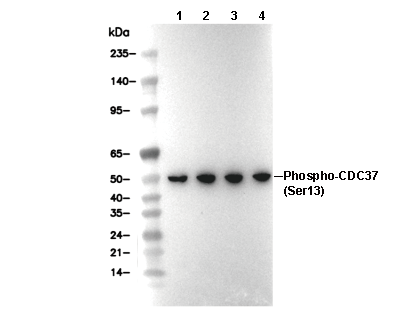 Lane 1: 293T, Lane 2: 3T3, Lane 3: C2C12, Lane 4: PC12