|
Hoe te citeren 1. Voor in-tekst citatie (materialen en methoden): 2. Voor de tabel met belangrijke bronnen: |
||
|
Gratis nummer: (877) 796-6397 -- Alleen VS en Canada -- |
Fax: +1-832-582-8590 Bestellingen: +1-832-582-8158 |
Technische ondersteuning: +1-832-582-8158 Ext:3 Gelieve uw bestelnummer in de e-mail te vermelden. Wij streven ernaar alle e-mailvragen binnen één werkdag te beantwoorden. |
Biologische Beschrijving
| Specificiteit | MMP12 Antibody [B22A15] herkent endogene niveaus van totaal MMP12-eiwit. |
|---|---|
| Achtergrond | Matrix-metalloproteïnase-12 (MMP-12) is een zinkafhankelijk endopeptidase dat een cruciale rol speelt bij de remodellering van de extracellulaire matrix (ECM) en ontsteking. Afgescheiden als een inactief zymogeen, wordt MMP-12 geactiveerd door ontstekingssignalen. Het katalytische domein bestaat uit drie α-helices, vijf β-sheets en een zinkbindingsplaats (His218/222/228), essentieel voor het klieven van ECM-componenten zoals elastine, collageen IV, laminine en fibronectine. Dit stelt MMP-12 in staat om weefselremodellering te mediëren tijdens verschillende pathologische processen. MMP-12 is betrokken bij meerdere ziekten, waaronder COPD en emfyseem, waar het de afbraak van elastine aanstuurt en bijdraagt aan longweefselvernietiging. Bij atherosclerose destabiliseert het plaques door de slagaderwand te verzwakken. Tijdens een ischemische beroerte verstoort MMP-12 de bloed-hersenbarrière door afbraak van tight junction-eiwitten zoals claudine-5 en occludine, wat neuronale schade verergert. Het moduleert ook immuunresponsen door TNF-α af te scheiden en macrofagen en neutrofielen te rekruteren. Terwijl MMP-12 nierschade bij snel progressieve glomerulonefritis verergert door ECM-afbraak en oxidatieve stress, kan het fibrose verminderen in modellen van lever- en longschade door MMP-2 en MMP-13 te onderdrukken. Bovendien draagt MMP-12 bij aan vasculaire schade door te trafficken naar de celkern van gladde spiercellen, wat duidt op een transcriptionele regulatierol, en het bindt aan fosfolipidemembranen via basische residuen (Arg117/127), wat snelle pericellulaire proteolyse vergemakkelijkt. |
Gebruiksinformatie
| Toepassing | WB, IP | Verdunning |
|
||||
|---|---|---|---|---|---|---|---|
| Reactiviteit | Human | ||||||
| Bron | Rabbit Monoclonal Antibody | MW | 54 kDa | ||||
| Opslagbuffer | PBS, pH 7.2+50% Glycerol+0.05% BSA+0.01% NaN3 | Opslag (Vanaf de datum van ontvangst) |
-20°C (avoid freeze-thaw cycles), 2 years | ||||
| WB |
Experimental Protocol:
Sample preparation
1. Tissue: Lyse the tissue sample by adding an appropriate volume of ice-cold RIPA/NP-40 Lysis Buffer (containing Protease Inhibitor Cocktail),and homogenize the tissue at a low temperature or lyse it by sonication on ice, then incubate on ice for 30 minutes. 2. Adherent cell: Aspirate the culture medium and transfer the cells into an EP tube. Wash the cells with ice-cold PBS twice. Add an appropriate volume of RIPA/NP-40 Lysis Buffer (containing Protease Inhibitor Cocktail), sonicate to lyse the cells, and incubate on ice for 30 minutes. 3. Suspension cell: Transfer the culture medium to a pre-cooled centrifuge tube. Centrifuge and aspirate the supernatant. Wash the cells with ice-cold PBS twice.Add an appropriate volume of RIPA/NP-40 Lysis Buffer (containing Protease Inhibitor Cocktail), sonicate to lyse the cells, and incubate on ice for 30 minutes. 4. Place the lysate into a pre-cooled microcentrifuge tube. Centrifuge at 4°C for 15 min. Collect the supernatant;
5. Remove a small volume of lysate to determine the protein concentration;
6. Combine the lysate with protein loading buffer. Boil 20 µL sample under 95-100°C for 5 min. Centrifuge for 5 min after cool down on ice.
Electrophoretic separation
1. According to the concentration of extracted protein, load appropriate amount of protein sample and marker onto SDS-PAGE gels for electrophoresis. Recommended separating gel (lower gel) concentration: 10%. Reference Table for Selecting SDS-PAGE Separation Gel Concentrations 2. Power up 80V for 30 minutes. Then the power supply is adjusted (110 V~150 V), the Marker is observed, and the electrophoresis can be stopped when the indicator band of the predyed protein Marker where the protein is located is properly separated. (Note that the current should not be too large when electrophoresis, too large current (more than 150 mA) will cause the temperature to rise, affecting the result of running glue. If high currents cannot be avoided, an ice bath can be used to cool the bath.)
Transfer membrane
1. Take out the converter, soak the clip and consumables in the pre-cooled converter;
2. Activate PVDF membrane with methanol for 1 min and rinse with transfer buffer;
3. Install it in the order of "black edge of clip - sponge - filter paper - filter paper - glue -PVDF membrane - filter paper - filter paper - sponge - white edge of clip"; 4. The protein was electrotransferred to PVDF membrane. ( 0.45 µm PVDF membrane is recommended ) Reference Table for Selecting PVDF Membrane Pore Size Specifications Recommended conditions for wet transfer: 200 mA, 120 min. ( Note that the transfer conditions can be adjusted according to the protein size. For high-molecular-weight proteins, a higher current and longer transfer time are recommended. However, ensure that the transfer tank remains at a low temperature to prevent gel melting.)
Block
1. After electrotransfer, wash the film with TBST at room temperature for 5 minutes;
2. Incubate the film in the blocking solution for 1 hour at room temperature;
3. Wash the film with TBST for 3 times, 5 minutes each time.
Antibody incubation
1. Use 5% skim milk powder to prepare the primary antibody working liquid (recommended dilution ratio for primary antibody 1:10000), gently shake and incubate with the film at 4°C overnight; 2. Wash the film with TBST 3 times, 5 minutes each time;
3. Add the secondary antibody to the blocking solution and incubate with the film gently at room temperature for 1 hour;
4. After incubation, wash the film with TBST 3 times for 5 minutes each time.
Antibody staining
1389. Add the prepared ECL luminescent substrate (or select other color developing substrate according to the second antibody) and mix evenly;
2. Incubate with the film for 1 minute, remove excess substrate (keep the film moist), wrap with plastic film, and expose in the imaging system. (Exposure time of at least 60s is recommended)
|
Referenties
|
Toepassingsgegevens
WB
Gevalideerd door Selleck
-
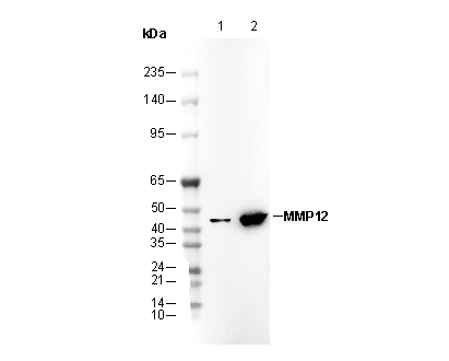 Lane 1: HT-1080, Lane 2: WI-38