|
Hoe te citeren 1. Voor in-tekst citatie (materialen en methoden): 2. Voor de tabel met belangrijke bronnen: |
||
|
Gratis nummer: (877) 796-6397 -- Alleen VS en Canada -- |
Fax: +1-832-582-8590 Bestellingen: +1-832-582-8158 |
Technische ondersteuning: +1-832-582-8158 Ext:3 Gelieve uw bestelnummer in de e-mail te vermelden. Wij streven ernaar alle e-mailvragen binnen één werkdag te beantwoorden. |
Biologische Beschrijving
| Specificiteit | Mitofusin 1 Antibody [D5A17] detecteert endogene niveaus van totaal Mitofusin-1 eiwit. |
|---|---|
| Achtergrond | Mitofusin 1 (MFN1) is een sleutellid van de dynamine-gerelateerde GTPase-familie, gelegen op de buitenste mitochondriale membraan, waar het essentieel is voor mitochondriale fusie, een proces dat van vitaal belang is voor het handhaven van de integriteit van het mitochondriale netwerk, energiehomeostase en celoverleving. MFN1 bevat een N-terminaal GTPase-domein dat GTP-binding en -hydrolyse mogelijk maakt, twee heptade-herhalingsgebieden (HR1 en HR2) die coiled-coil-structuren vormen die dimerisatie en tethering tussen aangrenzende mitochondriën mediëren, en twee transmembraanhelices die het eiwit in de buitenste membraan verankeren. De uitgebreide HR2 vergemakkelijkt directe interactie met MFN1 of MFN2 op naburige mitochondriën om efficiënte fusie te stimuleren. MFN1 werkt nauw samen met MFN2 en OPA1 om de mitochondriale morfologie te behouden, mitochondriaal DNA (mtDNA) te stabiliseren, ATP-productie te reguleren, apoptosewegen te ondersteunen en metabole aanpassing onder stress mogelijk te maken. Het bevordert ook mitofagie, waardoor de accumulatie van disfunctionele mitochondriën wordt voorkomen, en is belangrijk voor embryogenese en neuronale ontwikkeling. Verlies of disfunctie van MFN1 schaadt de mitochondriale dynamiek, wat energietekorten, neurodegeneratie en metabole ziekten veroorzaakt. Hoewel MFN1-mutaties minder vaak voorkomen dan die in MFN2, verergert deficiëntie de Charcot-Marie-Tooth type 2A-achtige neuropathie, spieratrofie en neuro-inflammatoire aandoeningen; veranderde MFN1-expressie wordt waargenomen bij ziekten zoals Parkinson, Alzheimer en sommige kankers. |
Gebruiksinformatie
| Toepassing | WB, IP | Verdunning |
|
||||
|---|---|---|---|---|---|---|---|
| Reactiviteit | Mouse, Rat | ||||||
| Bron | Rabbit Monoclonal Antibody | MW | 84 kDa | ||||
| Opslagbuffer | PBS, pH 7.2+50% Glycerol+0.05% BSA+0.01% NaN3 | Opslag (Vanaf de datum van ontvangst) |
-20°C (avoid freeze-thaw cycles), 2 years | ||||
| WB |
Experimental Protocol:
Sample preparation
1. Tissue: Lyse the tissue sample by adding an appropriate volume of ice-cold RIPA/NP-40 Lysis Buffer (containing Protease Inhibitor Cocktail),and homogenize the tissue at a low temperature. 2. Adherent cell: Aspirate the culture medium and wash the cells with ice-cold PBS twice. Lyse the cells by adding an appropriate volume of RIPA/NP-40 Lysis Buffer (containing Protease Inhibitor Cocktail) and put the sample on ice for 5 min. 3. Suspension cell: Transfer the culture medium to a pre-cooled centrifuge tube. Centrifuge and aspirate the supernatant. Wash the cells with ice-cold PBS twice. Lyse the cells by adding an appropriate volume of RIPA/NP-40 Lysis Buffer (containing Protease Inhibitor Cocktail) and put the sample on ice for 5 min. 4. Place the lysate into a pre-cooled microcentrifuge tube. Centrifuge at 4°C for 15 min. Collect the supernatant;
5. Remove a small volume of lysate to determine the protein concentration;
6. Combine the lysate with protein loading buffer. Boil 20 µL sample under 95-100°C for 5 min. Centrifuge for 5 min after cool down on ice.
Electrophoretic separation
1. According to the concentration of extracted protein, load appropriate amount of protein sample and marker onto SDS-PAGE gels for electrophoresis. Recommended separating gel (lower gel) concentration: 10%. Reference Table for Selecting SDS-PAGE Separation Gel Concentrations 2. Power up 80V for 30 minutes. Then the power supply is adjusted (110 V~150 V), the Marker is observed, and the electrophoresis can be stopped when the indicator band of the predyed protein Marker where the protein is located is properly separated. (Note that the current should not be too large when electrophoresis, too large current (more than 150 mA) will cause the temperature to rise, affecting the result of running glue. If high currents cannot be avoided, an ice bath can be used to cool the bath.)
Transfer membrane
1. Take out the converter, soak the clip and consumables in the pre-cooled converter;
2. Activate PVDF membrane with methanol for 1 min and rinse with transfer buffer;
3. Install it in the order of "black edge of clip - sponge - filter paper - filter paper - glue -PVDF membrane - filter paper - filter paper - sponge - white edge of clip"; 4. The protein was electrotransferred to PVDF membrane. ( 0.45 µm PVDF membrane is recommended ) Reference Table for Selecting PVDF Membrane Pore Size Specifications Recommended conditions for wet transfer: 200 mA, 120 min. ( Note that the transfer conditions can be adjusted according to the protein size. For high-molecular-weight proteins, a higher current and longer transfer time are recommended. However, ensure that the transfer tank remains at a low temperature to prevent gel melting.)
Block
1. After electrotransfer, wash the film with TBST at room temperature for 5 minutes;
2. Incubate the film in the blocking solution for 1 hour at room temperature;
3. Wash the film with TBST for 3 times, 5 minutes each time.
Antibody incubation
1. Use 5% skim milk powder to prepare the primary antibody working liquid (recommended dilution ratio for primary antibody 1:1000), gently shake and incubate with the film at 4°C overnight; 2. Wash the film with TBST 3 times, 5 minutes each time;
3. Add the secondary antibody to the blocking solution and incubate with the film gently at room temperature for 1 hour;
4. After incubation, wash the film with TBST 3 times for 5 minutes each time.
Antibody staining
1. Add the prepared ECL luminescent substrate (or select other color developing substrate according to the second antibody) and mix evenly;
2. Incubate with the film for 1 minute, remove excess substrate (keep the film moist), wrap with plastic film, and expose in the imaging system.
|
Referenties
|
Toepassingsgegevens
WB
Gevalideerd door Selleck
-
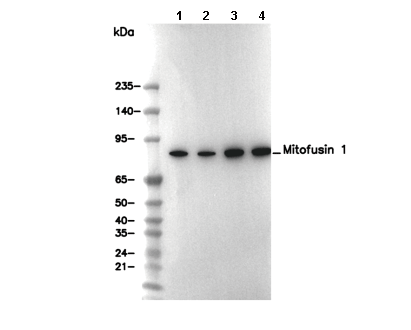 Lane 1: NIH/3T3, Lane 2: Neuro-2a, Lane 3: Mouse brain, Lane 4: Rat brain