|
Hoe te citeren 1. Voor in-tekst citatie (materialen en methoden): 2. Voor de tabel met belangrijke bronnen: |
||
|
Gratis nummer: (877) 796-6397 -- Alleen VS en Canada -- |
Fax: +1-832-582-8590 Bestellingen: +1-832-582-8158 |
Technische ondersteuning: +1-832-582-8158 Ext:3 Gelieve uw bestelnummer in de e-mail te vermelden. Wij streven ernaar alle e-mailvragen binnen één werkdag te beantwoorden. |
Biologische Beschrijving
| Specificiteit | INPP4b Antibody [J17B9] herkent endogene niveaus van totaal INPP4b-eiwit. |
|---|---|
| Achtergrond | Inositolpolyfosfaat 4-fosfatase type II (INPP4B) is een opkomend kankergen met een dubbele rol, voornamelijk erkend voor zijn rol als negatieve regulator van de fosfo-inositide 3-kinase (PI3K)/AKT-signaalroute. INPP4B wordt wijd tot expressie gebracht in menselijke weefsels, met de hoogste niveaus gevonden in het hart en skeletspieren. Het INPP4B-gen is significant geassocieerd met de variabiliteit van de botmassadichtheid bij mensen en is geïdentificeerd als een locus voor osteoporosegevoeligheid. INPP4B speelt ook een rol bij de differentiatie van mannelijke kiemcellen en de testiculaire fysiologie, waarbij de expressie ervan opmerkelijk toeneemt in post-meiotische kiemcellen van mannelijke muizen en mensen tijdens de spermatogenese. INPP4B is diffuus verspreid door het cytosol en transloceert bij groeifactorstimulatie naar het plasmamembraan om PtdIns(3,4)P2-gemedieerde AKT-activering negatief te reguleren. De fosfo-eiwitdoelen van INPP4B omvatten fosfo-Tyr-residuen op AKT1 en fosfo-Ser380- en -Thr382/383-residuen op PTEN, wat aangeeft dat INPP4B de PI3K/AKT-signalering kan reguleren via zowel zijn lipide- als eiwitfosfatase-activiteiten. |
Gebruiksinformatie
| Toepassing | WB, IP, IHC | Verdunning |
|
||||||
|---|---|---|---|---|---|---|---|---|---|
| Reactiviteit | Human | ||||||||
| Bron | Rabbit Monoclonal Antibody | MW | 110 kda | ||||||
| Opslagbuffer | PBS, pH 7.2+50% Glycerol+0.05% BSA+0.01% NaN₃ | Opslag (Vanaf de datum van ontvangst) |
–20°C (avoid freeze-thaw cycles), 2 years | ||||||
| WB |
Experimental Protocol:
Sample preparation
1. Tissue: Lyse the tissue sample by adding an appropriate volume of ice-cold RIPA/NP-40 Lysis Buffer (containing Protease Inhibitor Cocktail),and homogenize the tissue at a low temperature or lyse it by sonication on ice, then incubate on ice for 30 minutes. 2. Adherent cell: Aspirate the culture medium and transfer the cells into an EP tube. Wash the cells with ice-cold PBS twice. Add an appropriate volume of RIPA/NP-40 Lysis Buffer (containing Protease Inhibitor Cocktail), sonicate to lyse the cells, and incubate on ice for 30 minutes. 3. Suspension cell: Transfer the culture medium to a pre-cooled centrifuge tube. Centrifuge and aspirate the supernatant. Wash the cells with ice-cold PBS twice.Add an appropriate volume of RIPA/NP-40 Lysis Buffer (containing Protease Inhibitor Cocktail), sonicate to lyse the cells, and incubate on ice for 30 minutes. 4. Place the lysate into a pre-cooled microcentrifuge tube. Centrifuge at 4°C for 15 min. Collect the supernatant;
5. Remove a small volume of lysate to determine the protein concentration;
6. Combine the lysate with protein loading buffer. Boil 20 µL sample under 95-100°C for 5 min. Centrifuge for 5 min after cool down on ice.
Electrophoretic separation
1. According to the concentration of extracted protein, load appropriate amount of protein sample and marker onto SDS-PAGE gels for electrophoresis. Recommended separating gel (lower gel) concentration: 5%. Reference Table for Selecting SDS-PAGE Separation Gel Concentrations 2. Power up 80V for 30 minutes. Then the power supply is adjusted (110 V~150 V), the Marker is observed, and the electrophoresis can be stopped when the indicator band of the predyed protein Marker where the protein is located is properly separated. (Note that the current should not be too large when electrophoresis, too large current (more than 150 mA) will cause the temperature to rise, affecting the result of running glue. If high currents cannot be avoided, an ice bath can be used to cool the bath.)
Transfer membrane
1. Take out the converter, soak the clip and consumables in the pre-cooled converter;
2. Activate PVDF membrane with methanol for 1 min and rinse with transfer buffer;
3. Install it in the order of "black edge of clip - sponge - filter paper - filter paper - glue -PVDF membrane - filter paper - filter paper - sponge - white edge of clip"; 4. The protein was electrotransferred to PVDF membrane. ( 0.45 µm PVDF membrane is recommended ) Reference Table for Selecting PVDF Membrane Pore Size Specifications Recommended conditions for wet transfer: 200 mA, 120 min. ( Note that the transfer conditions can be adjusted according to the protein size. For high-molecular-weight proteins, a higher current and longer transfer time are recommended. However, ensure that the transfer tank remains at a low temperature to prevent gel melting.)
Block
1. After electrotransfer, wash the film with TBST at room temperature for 5 minutes;
2. Incubate the film in the blocking solution for 1 hour at room temperature;
3. Wash the film with TBST for 3 times, 5 minutes each time.
Antibody incubation
1. Use 5% skim milk powder to prepare the primary antibody working liquid (recommended dilution ratio for primary antibody 1:1000), gently shake and incubate with the film at 4°C overnight; 2. Wash the film with TBST 3 times, 5 minutes each time;
3. Add the secondary antibody to the blocking solution and incubate with the film gently at room temperature for 1 hour;
4. After incubation, wash the film with TBST 3 times for 5 minutes each time.
Antibody staining
683. Add the prepared ECL luminescent substrate (or select other color developing substrate according to the second antibody) and mix evenly;
2. Incubate with the film for 1 minute, remove excess substrate (keep the film moist), wrap with plastic film, and expose in the imaging system.
|
| IHC |
Experimental Protocol: Deparaffinization/Rehydration
1. Deparaffinize/hydrate sections:
2. Incubate sections in three washes of xylene for 5 min each.
3. Incubate sections in two washes of 100% ethanol for 10 min each.
4. Incubate sections in two washes of 95% ethanol for 10 min each.
5. Wash sections two times in dH2O for 5 min each.
6.Antigen retrieval: For Citrate: Heat slides in a microwave submersed in 1X citrate unmasking solution until boiling is initiated; continue with 10 min at a sub-boiling temperature (95°-98°C). Cool slides on bench top for 30 min.
Staining
1. Wash sections in dH2O three times for 5 min each.
2. Incubate sections in 3% hydrogen peroxide for 10 min.
3. Wash sections in dH2O two times for 5 min each.
4. Wash sections in wash buffer for 5 min.
5. Block each section with 100–400 µl of blocking solution for 1 hr at room temperature.
6. Remove blocking solution and add 100–400 µl primary antibody diluent in to each section. Incubate overnight at 4°C.
7. Remove antibody solution and wash sections with wash buffer three times for 5 min each.
8. Cover section with 1–3 drops HRPas needed. Incubate in a humidified chamber for 30 min at room temperature.
9. Wash sections three times with wash buffer for 5 min each.
10. Add DAB Chromogen Concentrate to DAB Diluent and mix well before use.
11. Apply 100–400 µl DAB to each section and monitor closely. 1–10 min generally provides an acceptable staining intensity.
12. Immerse slides in dH2O.
13. If desired, counterstain sections with hematoxylin.
14. Wash sections in dH2O two times for 5 min each.
15. Dehydrate sections: Incubate sections in 95% ethanol two times for 10 sec each; Repeat in 100% ethanol, incubating sections two times for 10 sec each; Repeat in xylene, incubating sections two times for 10 sec each.
16. Mount sections with coverslips and mounting medium.
|
Referenties
|
Toepassingsgegevens
WB
Gevalideerd door Selleck
-
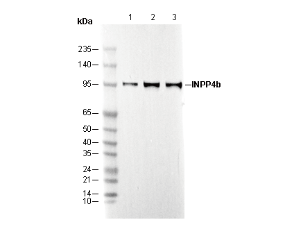 Lane 1: MCF7
Lane 2: A-204
Lane 3: ZR-75-1
IHC
Gevalideerd door Selleck
-
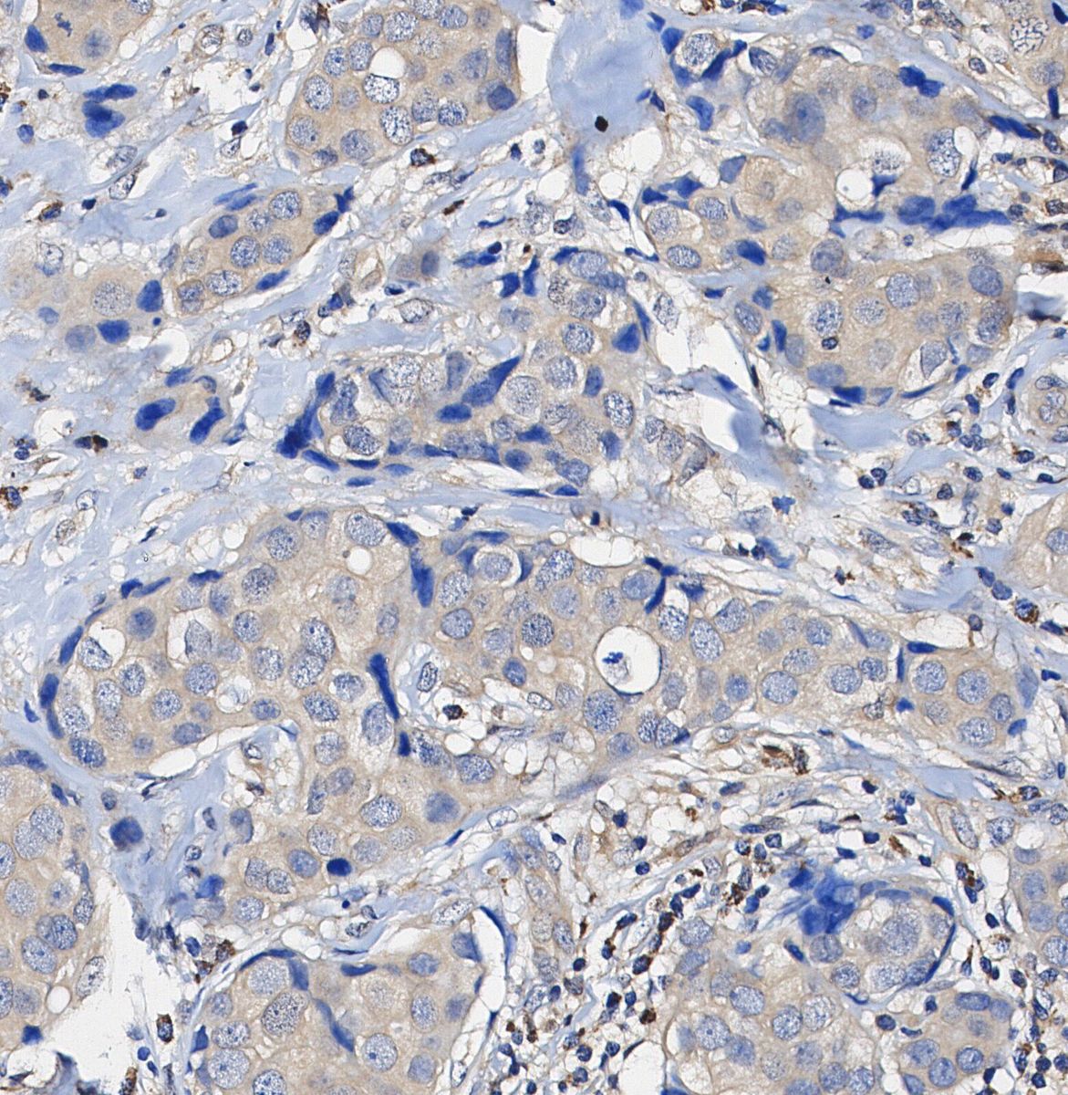 Immunohistochemical analysis of formalin fixed paraffin embedded human Colorectal cancer tissue with F1415 at 1/100 dilution.