|
Hoe te citeren 1. Voor in-tekst citatie (materialen en methoden): 2. Voor de tabel met belangrijke bronnen: |
||
|
Gratis nummer: (877) 796-6397 -- Alleen VS en Canada -- |
Fax: +1-832-582-8590 Bestellingen: +1-832-582-8158 |
Technische ondersteuning: +1-832-582-8158 Ext:3 Gelieve uw bestelnummer in de e-mail te vermelden. Wij streven ernaar alle e-mailvragen binnen één werkdag te beantwoorden. |
Biologische Beschrijving
| Specificiteit | CA19-9 Antibody [C17A13] detecteert endogene niveaus van CA 19-9 eiwit. |
|---|---|
| Achtergrond | CA19-9 (koolhydraatantigeen 19-9), ook bekend als Sialyl Lewis A (sLeᵃ), is een gesialyleerd Lewis-bloedgroepantigeen, een tetrasaccharide glycaanepitoop bestaande uit sialinezuur, galactose, N-acetylglucosamine en fucose, gebiosynthetiseerd via de Lewis-bloedgroeppathway en vereist een functioneel Lewis-antigeen voor de vorming ervan. CA19-9 wordt tot expressie gebracht op glycoproteïnen en glycolipiden van epitheelcelmembranen, met name in de alvleesklier, galwegen, maag en dikke darm, en kan in de bloedbaan terechtkomen. Het is de huidige gouden standaard serumbiomarker voor pancreasadenocarcinoom. Functioneel dient CA19-9 als biomarker (voor diagnose, prognose, monitoring van de behandelingsrespons en detectie van recidieven), een voorspeller (correleert met tumorlast, stadium en reseceerbaarheid) en een promotor van kankerprogressie door E-selectine-gemedieerde adhesie te vergemakkelijken, angiogenese te verbeteren, immuunresponsen te moduleren en interacties in de tumormicro-omgeving te beïnvloeden. Naast pancreaskanker is CA19-9 verhoogd bij andere gastro-intestinale maligniteiten en bepaalde goedaardige ziekten, en wordt het onderzocht als therapeutisch doelwit via antilichamen, vaccins, biosyntheseremmers en CA19-9-geleide medicijnafgiftesystemen. |
Gebruiksinformatie
| Toepassing | IHC, FCM, ELISA | Verdunning |
|
||
|---|---|---|---|---|---|
| Reactiviteit | Human | ||||
| Bron | Mouse Monoclonal Antibody | MW | |||
| Opslagbuffer | PBS, pH 7.2+50% Glycerol+0.05% BSA+0.01% NaN3 | Opslag (Vanaf de datum van ontvangst) |
-20°C (avoid freeze-thaw cycles), 2 years | ||
| IHC |
Experimental Protocol:
Deparaffinization/Rehydration
1. Deparaffinize/hydrate sections:
2. Incubate sections in three washes of xylene for 5 min each.
3. Incubate sections in two washes of 100% ethanol for 10 min each.
4. Incubate sections in two washes of 95% ethanol for 10 min each.
5. Wash sections two times in dH2O for 5 min each.
6.Antigen retrieval: For Citrate: Heat slides in a microwave submersed in 1X citrate unmasking solution until boiling is initiated; continue with 10 min at a sub-boiling temperature (95°-98°C). Cool slides on bench top for 30 min.
Staining
1. Wash sections in dH2O three times for 5 min each.
2. Incubate sections in 3% hydrogen peroxide for 10 min.
3. Wash sections in dH2O two times for 5 min each.
4. Wash sections in wash buffer for 5 min.
5. Block each section with 100–400 µl of blocking solution for 1 hr at room temperature.
6. Remove blocking solution and add 100–400 µl primary antibody diluent in to each section. Incubate overnight at 4°C.
7. Remove antibody solution and wash sections with wash buffer three times for 5 min each.
8. Cover section with 1–3 drops HRPas needed. Incubate in a humidified chamber for 30 min at room temperature.
9. Wash sections three times with wash buffer for 5 min each.
10. Add DAB Chromogen Concentrate to DAB Diluent and mix well before use.
11. Apply 100–400 µl DAB to each section and monitor closely. 1–10 min generally provides an acceptable staining intensity.
12. Immerse slides in dH2O.
13. If desired, counterstain sections with hematoxylin.
14. Wash sections in dH2O two times for 5 min each.
15. Dehydrate sections: Incubate sections in 95% ethanol two times for 10 sec each; Repeat in 100% ethanol, incubating sections two times for 10 sec each; Repeat in xylene, incubating sections two times for 10 sec each.
16. Mount sections with coverslips and mounting medium.
|
Referenties
|
Toepassingsgegevens
IHC
Gevalideerd door Selleck
-
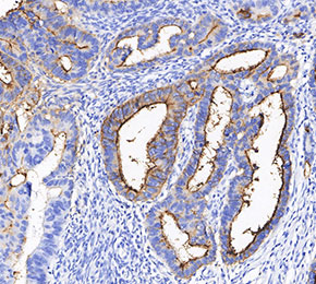 Immunohistochemical analysis of formalin fixed paraffin embedded human colorectal cancer tissue with F2466 at 1:100 dilution.